

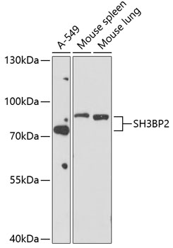
Anti-SH3BP2 Antibody
A12856
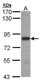
ApplicationsWestern Blot
Product group Antibodies
ReactivityHuman, Mouse, Rat
Overview
- SupplierAntibodies.com
- Product NameAnti-SH3BP2 Antibody
- Delivery Days Customer7
- ApplicationsWestern Blot
- CertificationResearch Use Only
- ClonalityPolyclonal
- ConjugateUnconjugated
- HostRabbit
- IsotypeIgG
- Scientific DescriptionRabbit polyclonal antibody to SH3BP2.
- ReactivityHuman, Mouse, Rat
- UNSPSC12352203
Related products
Product group Antibodies
SH3BP2 AntibodyCSB-PA021224LA01HU
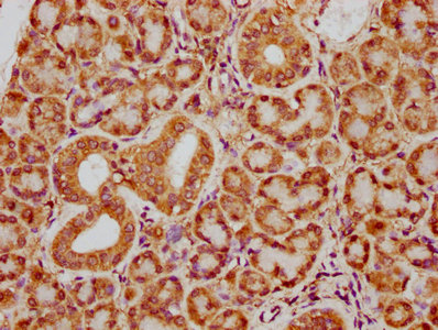
ApplicationsELISA, ImmunoHistoChemistry
ReactivityHuman
TargetSH3BP2
- SizePrice
Product group Antibodies
Anti-SH3BP2 Antibody144-61794
ApplicationsWestern Blot
ReactivityHuman, Mouse, Rat
TargetSH3BP2
- SizePrice

Product group Antibodies
Anti-SH3BP2 AntibodyHPA036789
ApplicationsImmunoCytoChemistry
ReactivityHuman
TargetSH3BP2
- SizePrice

Product group Antibodies
Anti-3BP2/SH3BP2 Antibody Picoband(r)A04008-1-CARRIER-FREE
ApplicationsFlow Cytometry, Western Blot, ELISA
ReactivityHuman
TargetSH3BP2
- SizePrice
Product group Antibodies

SH3BP2 AntibodyLS-C496951
ApplicationsWestern Blot
ReactivityHuman, Mouse, Rat
TargetSH3BP2
- SizePrice
Product group Antibodies
3BP2 antibodyGTX109708
ApplicationsWestern Blot
ReactivityHuman
TargetSH3BP2
- SizePrice