

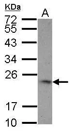
Sample (30 ug of whole cell lysate) A: HCT116 12% SDS PAGE GTX17355 diluted at 1:1000
![TNFSF9 antibody [N2C3] immunoprecipitates TNFSF9 protein in IP experiments. IP samples: A549 membrane extract A. Control with 4 μg of preimmune Rabbit IgG B. Immunoprecipitation of TNFSF9 protein by 4 μg TNFSF9 antibody [N2C3] (GTX117355) 15 % SDS-PAGE The immunoprecipitated TNFSF9 protein was detected by TNFSF9 antibody [N2C3] (GTX117355) diluted at 1:500. [EasyBlot anti-rabbit IgG (GTX221666-01) was used as a secondary reagent]](https://www.genetex.com/upload/website/prouct_img/normal/GTX117355/GTX117355_40555_IP_w_23060519_112.webp "TNFSF9 antibody [N2C3] immunoprecipitates TNFSF9 protein in IP experiments. IP samples: A549 membrane extract A. Control with 4 μg of preimmune Rabbit IgG B. Immunoprecipitation of TNFSF9 protein by 4 μg TNFSF9 antibody [N2C3] (GTX117355) 15 % SDS-PAGE The immunoprecipitated TNFSF9 protein was detected by TNFSF9 antibody [N2C3] (GTX117355) diluted at 1:500. [EasyBlot anti-rabbit IgG (GTX221666-01) was used as a secondary reagent]")
Sample (30 ug of whole cell lysate) A: HCT116 12% SDS PAGE GTX17355 diluted at 1:1000
4-1BBL / CD137L antibody [N2C3]
GTX117355
ApplicationsImmunoPrecipitation, Western Blot
Product group Antibodies
ReactivityHuman
TargetTNFSF9
Overview
- SupplierGeneTex
- Product Name4-1BBL / CD137L antibody [N2C3]
- Delivery Days Customer9
- Application Supplier NoteWB: 1:500-1:3000. IP: 1:100-1:500. *Optimal dilutions/concentrations should be determined by the researcher.Not tested in other applications.
- ApplicationsImmunoPrecipitation, Western Blot
- CertificationResearch Use Only
- ClonalityPolyclonal
- Concentration1 mg/ml
- ConjugateUnconjugated
- Gene ID8744
- Target nameTNFSF9
- Target descriptionTNF superfamily member 9
- Target synonyms4-1BB-L, CD137L, TNLG5A, tumor necrosis factor ligand superfamily member 9, 4-1BB ligand, 4-1BBL, CD137 ligand, homolog of mouse 4-1BB-L, receptor 4-1BB ligand, tumor necrosis factor (ligand) superfamily, member 9, tumor necrosis factor ligand 5A, tumor necrosis factor superfamily member 9
- HostRabbit
- IsotypeIgG
- Protein IDP41273
- Protein NameTumor necrosis factor ligand superfamily member 9
- Scientific DescriptionThe protein encoded by this gene is a cytokine that belongs to the tumor necrosis factor (TNF) ligand family. This transmembrane cytokine is a bidirectional signal transducer that acts as a ligand for TNFRSF9/4-1BB, which is a costimulatory receptor molecule in T lymphocytes. This cytokine and its receptor are involved in the antigen presentation process and in the generation of cytotoxic T cells. The receptor TNFRSF9/4-1BB is absent from resting T lymphocytes but rapidly expressed upon antigenic stimulation. The ligand encoded by this gene, TNFSF9/4-1BBL, has been shown to reactivate anergic T lymphocytes in addition to promoting T lymphocyte proliferation. This cytokine has also been shown to be required for the optimal CD8 responses in CD8 T cells. This cytokine is expressed in carcinoma cell lines, and is thought to be involved in T cell-tumor cell interaction.
- ReactivityHuman
- Storage Instruction-20°C or -80°C,2°C to 8°C
- UNSPSC41116161
Datasheet
Related products
Product group Antibodies
Anti-TNFSF9 AntibodyA99503
ApplicationsImmunoFluorescence, Western Blot, ELISA
ReactivityHuman
- SizePrice
Product group Antibodies
anti-CD137L (human), mAb (41B436)AG-20A-0031
ApplicationsFlow Cytometry, Western Blot, ELISA, ImmunoCytoChemistry
ReactivityHuman
TargetTNFSF9
- SizePrice

Product group Antibodies
Anti-4-1BBL/Tnfsf9 Antibody Picoband(r)A06032-2-CARRIER-FREE
ApplicationsFlow Cytometry, Western Blot, ELISA
ReactivityMouse, Rat
TargetTNFSF9
- SizePrice

Product group Antibodies
TNFSF9 Polyclonal AntibodyBS-3851R
ApplicationsImmunoFluorescence, Western Blot, ELISA, ImmunoCytoChemistry, ImmunoHistoChemistry, ImmunoHistoChemistry Frozen, ImmunoHistoChemistry Paraffin
ReactivityHuman, Mouse, Rat
TargetTNFSF9
- SizePrice
Product group Antibodies
TNFSF9 AntibodyCSB-PA001424
ApplicationsImmunoFluorescence, ELISA
ReactivityHuman
TargetTNFSF9
- SizePrice
Product group Antibodies
ApplicationsImmunoPrecipitation, Western Blot, ImmunoCytoChemistry, ImmunoHistoChemistry
TargetTNFSF9
- SizePrice
Product group Antibodies

TNFSF9 / CD137L Antibody (PerCP)LS-C483115
ApplicationsFlow Cytometry
ReactivityMouse
TargetTNFSF9
- SizePrice

Product group Antibodies
Anti-TNFSF9 AntibodyHPA059857
ApplicationsImmunoCytoChemistry
ReactivityHuman
TargetTNFSF9
- SizePrice