

67kDa Laminin Receptor Antibody (YA1959)
HY-P82214
TargetRPSA
Product group Antibodies
Overview
- SupplierMedChem Express
- Product Name67kDa Laminin Receptor Antibody (YA1959)
- Delivery Days Customer5
- CertificationResearch Use Only
- ClonalityMonoclonal
- Gene ID3921
- Target nameRPSA
- Target descriptionribosomal protein SA
- Target synonyms37 kDa laminin receptor; 37/67 kDa laminin receptor; 37LRP; 40S ribosomal protein SA; 67 kDa laminin receptor; 67LR; colon carcinoma laminin-binding protein; ICAS; LAMBR; laminin receptor 1 (67kD, ribosomal protein SA); laminin-binding protein precursor p40; lamR; LAMR1; LBP; LBP/p40; LRP; LRP/LR; multidrug resistance-associated protein MGr1-Ag; NEM/1CHD4; p40; SA; small ribosomal subunit protein uS2
- HostRabbit
- IsotypeIgG
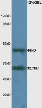
- Scientific Description67kDa Laminin Receptor Antibody (YA1959) is a rabbit-derived non-conjugated IgG antibody (Clone NO.: YA1959), targeting 67kDa Laminin Receptor, with a predicted molecular weight of 33 kDa (observed band size: 40 kDa). 67kDa Laminin Receptor Antibody (YA1959) can be used for WB, IP experiment in human, mouse, rat background.
- Storage Instruction-20°C
- UNSPSC12352203
Related products
Product group Antibodies
RPSA Polyclonal AntibodyCAC13914
ApplicationsWestern Blot, ELISA
TargetRPSA
- SizePrice
Product group Antibodies
References
RPSA Polyclonal AntibodyBS-0900R
ApplicationsFlow Cytometry, ImmunoFluorescence, Western Blot, ELISA, ImmunoCytoChemistry, ImmunoHistoChemistry, ImmunoHistoChemistry Frozen, ImmunoHistoChemistry Paraffin
TargetRPSA
- SizePrice
Product group Antibodies
RPSA antibody [N1C3]GTX100831
ApplicationsWestern Blot, ImmunoHistoChemistry, ImmunoHistoChemistry Paraffin
TargetRPSA
- SizePrice
Product group Antibodies
RPSA AntibodyCSB-PA009782
ApplicationsWestern Blot, ELISA, ImmunoHistoChemistry
TargetRPSA
- SizePrice
Product group Antibodies
ApplicationsImmunoPrecipitation, Western Blot
TargetRPSA
- SizePrice
Product group Antibodies
Anti-Mouse RPSA Antibody144-09008
ApplicationsWestern Blot
TargetRPSA
- SizePrice
Product group Antibodies

RPSA / Laminin Receptor AntibodyLS-C400882
ApplicationsWestern Blot, ELISA
TargetRPSA
- SizePrice

Product group Antibodies
Anti-67kDa Laminin Receptor/RPSA Antibody Picoband(r)PB10094-CARRIER-FREE
ApplicationsFlow Cytometry, ImmunoFluorescence, Western Blot, ImmunoCytoChemistry, ImmunoHistoChemistry, ImmunoHistoChemistry Frozen
TargetRPSA
- SizePrice