


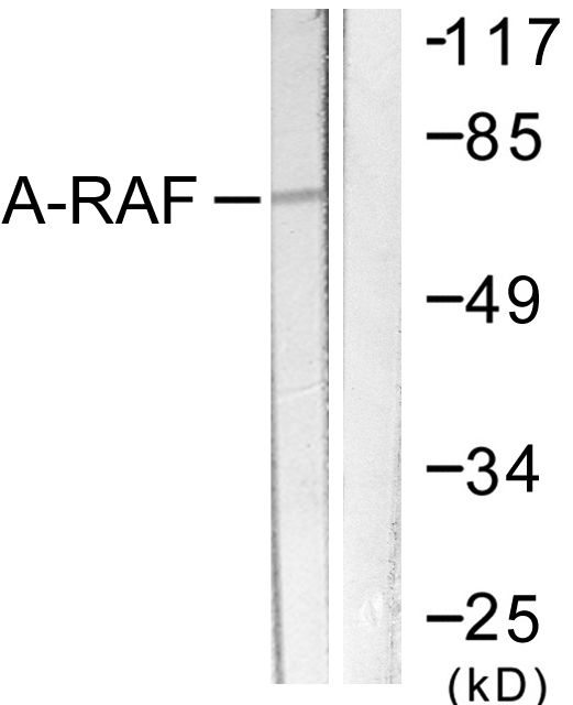
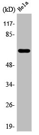
HeLa cell lysates probed with A-RAF (24D1) Monoclonal Antibody (bsm-52015R) at 1:1000 overnight at 4˚C. Followed by a conjugated secondary antibody.
 for 15min; Block endogenous peroxidase by 3% hydrogen peroxide for 30 minutes; Blocking buffer at 37°C for 20min; Antibody incubation with A-RAF (24D1) Monoclonal Antibody (bsm-52015R) at 1:50 overnight at 4°C, followed by a conjugated secondary and DAB staining.")
![NIH/3T3 cells were stained with A-RAF (24D1) Monoclonal Antibody (bsm-52015R) at [1:200] incubated overnight at 4C, followed by secondary antibody incubation, DAPI staining of the nuclei and detection.](https://biossantibodies.com/image-raw/16256.jpeg "NIH/3T3 cells were stained with A-RAF (24D1) Monoclonal Antibody (bsm-52015R) at [1:200] incubated overnight at 4C, followed by secondary antibody incubation, DAPI staining of the nuclei and detection.")
![HeLa cells were stained with A-RAF (24D1) Monoclonal Antibody (bsm-52015R) at [1:200] incubated overnight at 4C, followed by secondary antibody incubation, DAPI staining of the nuclei and detection.](https://biossantibodies.com/image-raw/16257.jpeg "HeLa cells were stained with A-RAF (24D1) Monoclonal Antibody (bsm-52015R) at [1:200] incubated overnight at 4C, followed by secondary antibody incubation, DAPI staining of the nuclei and detection.")
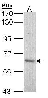
 at 1:1000 dilution and 4°C overnight incubation. Followed by conjugated secondary antibody incubation at 1:20000 for 60 min at 37˚C.")
HeLa cell lysates probed with A-RAF (24D1) Monoclonal Antibody (bsm-52015R) at 1:1000 overnight at 4˚C. Followed by a conjugated secondary antibody.
A-RAF Recombinant Antibody
BSM-52015R
ApplicationsFlow Cytometry, ImmunoFluorescence, Western Blot, ImmunoCytoChemistry, ImmunoHistoChemistry, ImmunoHistoChemistry Frozen, ImmunoHistoChemistry Paraffin
Product group Antibodies
ReactivityHuman
TargetARAF
Overview
- SupplierBioss
- Product NameA-RAF Recombinant Antibody
- Delivery Days Customer16
- ApplicationsFlow Cytometry, ImmunoFluorescence, Western Blot, ImmunoCytoChemistry, ImmunoHistoChemistry, ImmunoHistoChemistry Frozen, ImmunoHistoChemistry Paraffin
- Applications SupplierWB(1:300-5000), IHC-P(1:200-400), IHC-F(1:100-500), IF(), FCM(1ug/Test), ICC/IF(1:50-100)
- CertificationResearch Use Only
- ClonalityMonoclonal
- ConjugateUnconjugated
- Gene ID369
- Target nameARAF
- Target descriptionA-Raf proto-oncogene, serine/threonine kinase
- Target synonymsA-RAF, ARAF1, PKS2, RAFA1, serine/threonine-protein kinase A-Raf, A-Raf proto-oncogene serine/threonine-protein kinase, Oncogene ARAF1, Ras-binding protein DA-Raf, proto-oncogene A-Raf-1, proto-oncogene Pks, v-raf murine sarcoma 3611 viral oncogene homolog 1, v-raf murine sarcoma 3611 viral oncogene-like protein
- HostRabbit
- IsotypeIgG
- Protein IDP10398
- Protein NameSerine/threonine-protein kinase A-Raf
- ReactivityHuman
- Storage Instruction-20°C
- UNSPSC41116161
Datasheet
Related products
Product group Antibodies
Anti-A-RAF AntibodyA95936
ApplicationsWestern Blot, ELISA, ImmunoHistoChemistry
ReactivityHuman, Mouse, Rat
- SizePrice

Product group Antibodies
Anti-ARAF Antibody Picoband(r)A02061-2-CARRIER-FREE
ApplicationsFlow Cytometry, ImmunoFluorescence, Western Blot, ELISA, ImmunoCytoChemistry
ReactivityHuman, Mouse, Rat
TargetARAF
- SizePrice
Product group Antibodies
ARAF AntibodyCSB-PA000918
ApplicationsWestern Blot, ELISA, ImmunoHistoChemistry
ReactivityHuman, Mouse, Rat
TargetARAF
- SizePrice
Product group Antibodies
A-RAF antibody [C1C3]GTX101427
ApplicationsImmunoFluorescence, Western Blot, ImmunoCytoChemistry, ImmunoHistoChemistry, ImmunoHistoChemistry Paraffin
ReactivityHuman, Mouse
TargetARAF
- SizePrice
Product group Antibodies

ARAF / ARAF1 / A-RAF Antibody (Internal)LS-C358904
ApplicationsWestern Blot
ReactivityHuman
TargetARAF
- SizePrice

Product group Antibodies
Anti-ARAF AntibodyHPA066326
ApplicationsWestern Blot, ImmunoHistoChemistry
ReactivityHuman
TargetARAF
- SizePrice
Product group Antibodies
ApplicationsWestern Blot, ELISA, ImmunoHistoChemistry, ImmunoHistoChemistry Paraffin
ReactivityHuman
TargetARAF
- SizePrice