

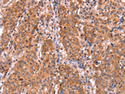
The image on the left is immunohistochemistry of paraffin-embedded Human gastic cancer tissue using CSB-PA196653(ABI1 Antibody) at dilution 1/40, on the right is treated with synthetic peptide. (Original magnification: x200)
 at dilution 1/40, on the right is treated with synthetic peptide. (Original magnification: x200)")
The image on the left is immunohistochemistry of paraffin-embedded Human gastic cancer tissue using CSB-PA196653(ABI1 Antibody) at dilution 1/40, on the right is treated with synthetic peptide. (Original magnification: x200)
ABI1 Antibody
CSB-PA196653
ApplicationsELISA, ImmunoHistoChemistry
Product group Antibodies
ReactivityHuman, Mouse, Rat
TargetABI1
Overview
- SupplierCusabio
- Product NameABI1 Antibody
- Delivery Days Customer20
- ApplicationsELISA, ImmunoHistoChemistry
- CertificationResearch Use Only
- ClonalityPolyclonal
- ConjugateUnconjugated
- Gene ID10006
- Target nameABI1
- Target descriptionabl interactor 1
- Target synonymsABI-1, ABLBP4, E3B1, NAP1BP, SSH3BP, SSH3BP1, abl interactor 1, Abelson interactor 1, Abl-interactor protein 1 long, abl-binding protein 4, eps8 SH3 domain-binding protein, interactor protein AblBP4, nap1 binding protein, spectrin SH3 domain-binding protein 1
- HostRabbit
- IsotypeIgG
- Protein IDQ8IZP0
- Protein NameAbl interactor 1
- Scientific DescriptionThis gene encodes a member of the Abelson-interactor family of adaptor proteins. These proteins facilitate signal transduction as components of several multiprotein complexes, and regulate actin polymerization and cytoskeletal remodeling through interactions with Abelson tyrosine kinases. The encoded protein plays a role in macropinocytosis as a component of the WAVE2 complex, and also forms a complex with EPS8 and SOS1 that mediates signal transduction from Ras to Rac. This gene may play a role in the progression of several malignancies including melanoma, colon cancer and breast cancer, and a t(10;11) chromosomal translocation involving this gene and the MLL gene has been associated with acute myeloid leukemia. Alternatively spliced transcript variants encoding multiple isoforms have been observed for this gene, and a pseudogene of this gene is located on the long arm of chromosome 14
- ReactivityHuman, Mouse, Rat
- Storage Instruction-20°C or -80°C
- UNSPSC41116161
Related products
Product group Antibodies
Anti-ABI1 AntibodyA38765
ApplicationsWestern Blot, ImmunoHistoChemistry
ReactivityHuman, Mouse, Rat
- SizePrice
Product group Antibodies
Anti-ABI1 Antibody144-64089
ApplicationsWestern Blot
ReactivityHuman, Mouse
TargetABI1
- SizePrice
Product group Antibodies
ABI1 Polyclonal AntibodyBS-1961R
ApplicationsImmunoFluorescence, Western Blot, ELISA, ImmunoCytoChemistry, ImmunoHistoChemistry, ImmunoHistoChemistry Frozen, ImmunoHistoChemistry Paraffin
ReactivityCanine, Chicken, Equine, Human, Mouse, Rabbit, Rat
TargetABI1
- SizePrice
Product group Antibodies
ApplicationsImmunoPrecipitation, Western Blot, ImmunoCytoChemistry, ImmunoHistoChemistry
ReactivityMouse, Porcine, Rat
TargetABI1
- SizePrice
Product group Antibodies

ABI1 / SSH3BP1 AntibodyLS-C402089
ApplicationsELISA, ImmunoHistoChemistry
ReactivityHuman, Mouse, Rat
TargetABI1
- SizePrice

Product group Antibodies
Anti-ABI1 AntibodyHPA068407
ApplicationsImmunoCytoChemistry
ReactivityHuman
TargetABI1
- SizePrice
Product group Antibodies
SSH3BP1 antibodyGTX111478
ApplicationsWestern Blot
ReactivityHuman, Mouse
TargetABI1
- SizePrice

Product group Antibodies
Anti-SSH3BP1/ABI1 Antibody Picoband(r)PB9416-CARRIER-FREE
ApplicationsFlow Cytometry, ImmunoFluorescence, Western Blot, ImmunoCytoChemistry, ImmunoHistoChemistry
ReactivityHamster, Human, Mouse, Rat
TargetABI1
- SizePrice