

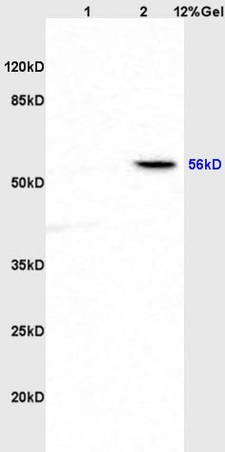
Lane 1: rat brain lysates Lane 2: rat heart lysates probed with Anti ABI1 Polyclonal Antibody, Unconjugated (bs-1961R) at 1:200 in 4˚C. Followed by conjugation to secondary antibody (bs-0295G-HRP) at 1:3000 90min in 37˚C. Predicted band 56kD. Observed band size: 56kD.
Lane 1: rat brain lysates Lane 2: rat heart lysates probed with Anti ABI1 Polyclonal Antibody, Unconjugated (bs-1961R) at 1:200 in 4˚C. Followed by conjugation to secondary antibody (bs-0295G-HRP) at 1:3000 90min in 37˚C. Predicted band 56kD. Observed band size: 56kD.
ABI1 Polyclonal Antibody
BS-1961R
ApplicationsImmunoFluorescence, Western Blot, ELISA, ImmunoCytoChemistry, ImmunoHistoChemistry, ImmunoHistoChemistry Frozen, ImmunoHistoChemistry Paraffin
Product group Antibodies
ReactivityCanine, Chicken, Equine, Human, Mouse, Rabbit, Rat
TargetABI1
Overview
- SupplierBioss
- Product NameABI1 Polyclonal Antibody
- Delivery Days Customer16
- ApplicationsImmunoFluorescence, Western Blot, ELISA, ImmunoCytoChemistry, ImmunoHistoChemistry, ImmunoHistoChemistry Frozen, ImmunoHistoChemistry Paraffin
- Applications SupplierWB(1:300-5000), ELISA(1:500-1000), IHC-P(1:200-400), IHC-F(1:100-500), IF(IHC-P)(1:50-200), IF(IHC-F)(1:50-200), IF(ICC)(1:50-200)
- CertificationResearch Use Only
- ClonalityPolyclonal
- Concentration1 ug/ul
- ConjugateUnconjugated
- Gene ID10006
- Target nameABI1
- Target descriptionabl interactor 1
- Target synonymsABI-1, ABLBP4, E3B1, NAP1BP, SSH3BP, SSH3BP1, abl interactor 1, Abelson interactor 1, Abl-interactor protein 1 long, abl-binding protein 4, eps8 SH3 domain-binding protein, interactor protein AblBP4, nap1 binding protein, spectrin SH3 domain-binding protein 1
- HostRabbit
- IsotypeIgG
- Protein IDQ8IZP0
- Protein NameAbl interactor 1
- ReactivityCanine, Chicken, Equine, Human, Mouse, Rabbit, Rat
- Storage Instruction-20°C
- UNSPSC41116161
Datasheet
Related products
Product group Antibodies
ABI1 AntibodyCSB-PA196653
ApplicationsELISA, ImmunoHistoChemistry
ReactivityHuman, Mouse, Rat
TargetABI1
- SizePrice
Product group Antibodies
ApplicationsImmunoPrecipitation, Western Blot, ImmunoCytoChemistry, ImmunoHistoChemistry
ReactivityMouse, Porcine, Rat
TargetABI1
- SizePrice
Product group Antibodies
Anti-ABI1 Antibody144-64089
ApplicationsWestern Blot
ReactivityHuman, Mouse
TargetABI1
- SizePrice
Product group Antibodies
Anti-ABI1 AntibodyA38765
ApplicationsWestern Blot, ImmunoHistoChemistry
ReactivityHuman, Mouse, Rat
- SizePrice

Product group Antibodies
Anti-ABI1 AntibodyHPA068407
ApplicationsImmunoCytoChemistry
ReactivityHuman
TargetABI1
- SizePrice
Product group Antibodies

ABI1 / SSH3BP1 AntibodyLS-C402089
ApplicationsELISA, ImmunoHistoChemistry
ReactivityHuman, Mouse, Rat
TargetABI1
- SizePrice

Product group Antibodies
Anti-SSH3BP1/ABI1 Antibody Picoband(r)PB9416-CARRIER-FREE
ApplicationsFlow Cytometry, ImmunoFluorescence, Western Blot, ImmunoCytoChemistry, ImmunoHistoChemistry
ReactivityHamster, Human, Mouse, Rat
TargetABI1
- SizePrice
Product group Antibodies
SSH3BP1 antibodyGTX111478
ApplicationsWestern Blot
ReactivityHuman, Mouse
TargetABI1
- SizePrice