

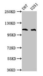
Western Blot Positive WB detected in: U87 whole cell lysate, U251 whole cell lysate All lanes: ABL2 antibody at 4microg/ml Secondary Goat polyclonal to rabbit IgG at 1/50000 dilution Predicted band size: 129, 125, 127, 115, 118, 116, 117, 61 kDa Observed band size: 115 kDa
Western Blot Positive WB detected in: U87 whole cell lysate, U251 whole cell lysate All lanes: ABL2 antibody at 4microg/ml Secondary Goat polyclonal to rabbit IgG at 1/50000 dilution Predicted band size: 129, 125, 127, 115, 118, 116, 117, 61 kDa Observed band size: 115 kDa
ABL2 Antibody
CSB-PA001106LA01HU
ApplicationsWestern Blot, ELISA
Product group Antibodies
ReactivityHuman
TargetABL2
Overview
- SupplierCusabio
- Product NameABL2 Antibody
- Delivery Days Customer20
- ApplicationsWestern Blot, ELISA
- CertificationResearch Use Only
- ClonalityPolyclonal
- ConjugateUnconjugated
- Gene ID27
- Target nameABL2
- Target descriptionABL proto-oncogene 2, non-receptor tyrosine kinase
- Target synonymsABLL, ARG, tyrosine-protein kinase ABL2, Abelson tyrosine-protein kinase 2, abelson-related gene protein, c-abl oncogene 2, non-receptor tyrosine kinase, tyrosine-protein kinase ARG, v-abl Abelson murine leukemia viral oncogene homolog 2
- HostRabbit
- IsotypeIgG
- Protein IDP42684
- Protein NameTyrosine-protein kinase ABL2
- Scientific DescriptionNon-receptor tyrosine-protein kinase that plays an ABL1-overlapping role in key processes linked to cell growth and survival such as cytoskeleton remodeling in response to extracellular stimuli, cell motility and adhesion and receptor endocytosis. Coordinates actin remodeling through tyrosine phosphorylation of proteins controlling cytoskeleton dynamics like MYH10 (involved in movement); CTTN (involved in signaling); or TUBA1 and TUBB (microtubule subunits). Binds directly F-actin and regulates actin cytoskeletal structure through its F-actin-bundling activity. Involved in the regulation of cell adhesion and motility through phosphorylation of key regulators of these processes such as CRK, CRKL, DOK1 or ARHGAP35. Adhesion-dependent phosphorylation of ARHGAP35 promotes its association with RASA1, resulting in recruitment of ARHGAP35 to the cell periphery where it inhibits RHO. Phosphorylates multiple receptor tyrosine kinases like PDGFRB and other substrates which are involved in endocytosis regulation such as RIN1. In brain, may regulate neurotransmission by phosphorylating proteins at the synapse. ABL2 acts also as a regulator of multiple pathological signaling cascades during infection. Pathogens can highjack ABL2 kinase signaling to reorganize the host actin cytoskeleton for multiple purposes, like facilitating intracellular movement and host cell exit. Finally, functions as its own regulator through autocatalytic activity as well as through phosphorylation of its inhibitor, ABI1.
- ReactivityHuman
- Storage Instruction-20°C or -80°C
- UNSPSC41116161
Related products
Product group Antibodies
Anti-ABL2 AntibodyA98304
ApplicationsWestern Blot, ELISA
ReactivityHuman, Mouse, Rat
- SizePrice
Product group Antibodies
Anti-ABL2 Antibody102-20129
ApplicationsWestern Blot, ImmunoHistoChemistry, ImmunoHistoChemistry Paraffin
TargetABL2
- SizePrice
Product group Antibodies
ABL2 Recombinant Antibody, Biotin ConjugatedBSM-61381R-BIOTIN
ApplicationsWestern Blot
ReactivityHuman, Mouse, Rat
TargetABL2
- SizePrice
Product group Antibodies
ABL2 Polyclonal AntibodyCAC15350
ApplicationsWestern Blot, ELISA
TargetABL2
- SizePrice
Product group Antibodies
ABL2 antibody [N1N3]GTX103584
ApplicationsWestern Blot
ReactivityHuman
TargetABL2
- SizePrice

Product group Antibodies
Anti-ABL2 AntibodyHPA072754
ApplicationsImmunoHistoChemistry
ReactivityHuman
TargetABL2
- SizePrice

Product group Antibodies
Anti-ABL2 Antibody Picoband(r)PB9913-CARRIER-FREE
ApplicationsWestern Blot, ImmunoHistoChemistry
ReactivityHuman, Mouse, Rat
TargetABL2
- SizePrice
Product group Antibodies

ABL2 AntibodyLS-C405850
ApplicationsWestern Blot, ELISA, ImmunoHistoChemistry
ReactivityHuman, Mouse
TargetABL2
- SizePrice