

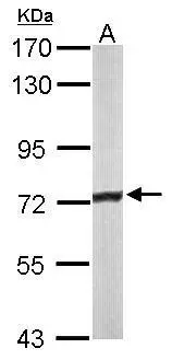
Sample (50 μg of whole cell lysate) A: mouse liver 7.5% SDS PAGE GTX114232 diluted at 1:3000 The HRP-conjugated anti-rabbit IgG antibody (GTX213110-01) was used to detect the primary antibody.
![ACADVL antibody [N1C1] detects ACADVL protein at cytoplasm by immunohistochemical analysis. Sample: Paraffin-embedded rat liver. ACADVL stained by ACADVL antibody [N1C1] (GTX114232) diluted at 1:500. Antigen Retrieval: Citrate buffer, pH 6.0, 15 min](https://www.genetex.com/upload/website/prouct_img/normal/GTX114232/GTX114232_43650_20200619_IHC-P_R_w_23060501_116.webp "ACADVL antibody [N1C1] detects ACADVL protein at cytoplasm by immunohistochemical analysis. Sample: Paraffin-embedded rat liver. ACADVL stained by ACADVL antibody [N1C1] (GTX114232) diluted at 1:500. Antigen Retrieval: Citrate buffer, pH 6.0, 15 min")
 antibody at 1:250 dilution.
Antigen Retrieval: Trilogy? (EDTA based, pH 8.0) buffer, 15min")
![ACADVL antibody [N1C1] detects ACADVL protein at mitochondria by immunohistochemical analysis. Sample: Paraffin-embedded human A549 xenograft. ACADVL antibody [N1C1] (GTX114232) diluted at 1:500.
Antigen Retrieval: Trilogy? (EDTA based, pH 8.0) buffer, 15min](https://www.genetex.com/upload/website/prouct_img/normal/GTX114232/GTX114232_40156_20151227_IHC-P_w_23060501_396.webp "ACADVL antibody [N1C1] detects ACADVL protein at mitochondria by immunohistochemical analysis. Sample: Paraffin-embedded human A549 xenograft. ACADVL antibody [N1C1] (GTX114232) diluted at 1:500.
Antigen Retrieval: Trilogy? (EDTA based, pH 8.0) buffer, 15min")
![ACADVL antibody [N1C1] detects ACADVL protein at cytoplasm by immunohistochemical analysis. Sample: Paraffin-embedded mouse kidney. ACADVL stained by ACADVL antibody [N1C1] (GTX114232) diluted at 1:500. Antigen Retrieval: Citrate buffer, pH 6.0, 15 min](https://www.genetex.com/upload/website/prouct_img/normal/GTX114232/GTX114232_43650_20200619_IHC-P_M_w_23060501_332.webp "ACADVL antibody [N1C1] detects ACADVL protein at cytoplasm by immunohistochemical analysis. Sample: Paraffin-embedded mouse kidney. ACADVL stained by ACADVL antibody [N1C1] (GTX114232) diluted at 1:500. Antigen Retrieval: Citrate buffer, pH 6.0, 15 min")
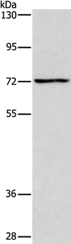
![Wild-type (WT) and ACADVL knockout (KO) 293T cell extracts (30 μg) were separated by 7.5% SDS-PAGE, and the membrane was blotted with ACADVL antibody [N1C1] (GTX114232) diluted at 1:1000. The HRP-conjugated anti-rabbit IgG antibody (GTX213110-01) was used to detect the primary antibody.](https://www.genetex.com/upload/website/prouct_img/normal/GTX114232/GTX114232_43649_20190802_WB_KO_watermark_w_23060501_692.webp "Wild-type (WT) and ACADVL knockout (KO) 293T cell extracts (30 μg) were separated by 7.5% SDS-PAGE, and the membrane was blotted with ACADVL antibody [N1C1] (GTX114232) diluted at 1:1000. The HRP-conjugated anti-rabbit IgG antibody (GTX213110-01) was used to detect the primary antibody.")
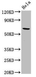
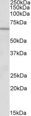
![Various whole cell extracts (30 μg) were separated by 7.5% SDS-PAGE, and the membrane was blotted with ACADVL antibody [N1C1] (GTX114232) diluted at 1:1000. The HRP-conjugated anti-rabbit IgG antibody (GTX213110-01) was used to detect the primary antibody. Corresponding RNA expression data for the same cell lines are based on Human Protein Atlas program.](https://www.genetex.com/upload/website/prouct_img/normal/GTX114232/GTX114232_43649_20191025_WB_TPM_watermark_w_23060501_478.webp "Various whole cell extracts (30 μg) were separated by 7.5% SDS-PAGE, and the membrane was blotted with ACADVL antibody [N1C1] (GTX114232) diluted at 1:1000. The HRP-conjugated anti-rabbit IgG antibody (GTX213110-01) was used to detect the primary antibody. Corresponding RNA expression data for the same cell lines are based on Human Protein Atlas program.")
![Various whole cell extracts (30 μg) were separated by 7.5% SDS-PAGE, and the membrane was blotted with ACADVL antibody [N1C1] (GTX114232) diluted at 1:1000. The HRP-conjugated anti-rabbit IgG antibody (GTX213110-01) was used to detect the primary antibody.](https://www.genetex.com/upload/website/prouct_img/normal/GTX114232/GTX114232_43649_20190802_WB_w_23060501_948.webp "Various whole cell extracts (30 μg) were separated by 7.5% SDS-PAGE, and the membrane was blotted with ACADVL antibody [N1C1] (GTX114232) diluted at 1:1000. The HRP-conjugated anti-rabbit IgG antibody (GTX213110-01) was used to detect the primary antibody.")
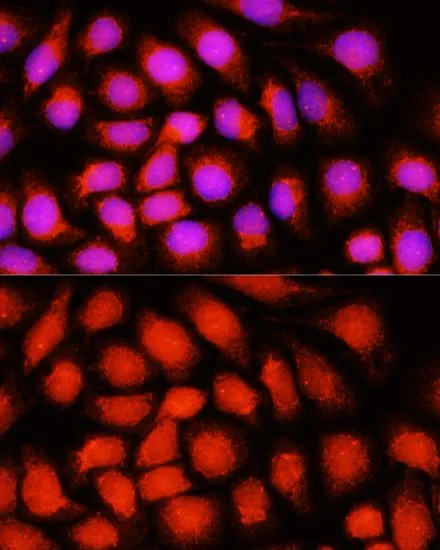
![ACADVL antibody [N1C1] detects ACADVL protein at mitochondria by immunofluorescent analysis. Sample: HeLa cells were fixed in ice-cold MeOH for 5 min. Green: ACADVL stained by ACADVL antibody [N1C1] (GTX114232) diluted at 1:500. Blue: Fluoroshield with DAPI (GTX30920). Scale bar= 10 μm.](https://www.genetex.com/upload/website/prouct_img/normal/GTX114232/GTX114232_43650_20200304_ICC_IF_w_23060501_701.webp "ACADVL antibody [N1C1] detects ACADVL protein at mitochondria by immunofluorescent analysis. Sample: HeLa cells were fixed in ice-cold MeOH for 5 min. Green: ACADVL stained by ACADVL antibody [N1C1] (GTX114232) diluted at 1:500. Blue: Fluoroshield with DAPI (GTX30920). Scale bar= 10 μm.")
Sample (50 μg of whole cell lysate) A: mouse liver 7.5% SDS PAGE GTX114232 diluted at 1:3000 The HRP-conjugated anti-rabbit IgG antibody (GTX213110-01) was used to detect the primary antibody.
ACADVL antibody [N1C1]
GTX114232
ApplicationsImmunoFluorescence, Western Blot, ImmunoCytoChemistry, ImmunoHistoChemistry, ImmunoHistoChemistry Paraffin
Product group Antibodies
ReactivityHuman, Mouse, Rat
TargetACADVL
Overview
- SupplierGeneTex
- Product NameACADVL antibody [N1C1]
- Delivery Days Customer9
- Application Supplier NoteWB: 1:500-1:3000. ICC/IF: 1:100-1:1000. IHC-P: 1:100-1:1000. *Optimal dilutions/concentrations should be determined by the researcher.Not tested in other applications.
- ApplicationsImmunoFluorescence, Western Blot, ImmunoCytoChemistry, ImmunoHistoChemistry, ImmunoHistoChemistry Paraffin
- CertificationResearch Use Only
- ClonalityPolyclonal
- Concentration0.3 mg/ml
- ConjugateUnconjugated
- Gene ID37
- Target nameACADVL
- Target descriptionacyl-CoA dehydrogenase very long chain
- Target synonymsACAD6, LCACD, VLCAD, very long-chain specific acyl-CoA dehydrogenase, mitochondrial, acyl-Coenzyme A dehydrogenase, very long chain
- HostRabbit
- IsotypeIgG
- Protein IDP49748
- Protein NameVery long-chain specific acyl-CoA dehydrogenase, mitochondrial
- Scientific DescriptionThe protein encoded by this gene is targeted to the inner mitochondrial membrane where it catalyzes the first step of the mitochondrial fatty acid beta-oxidation pathway. This acyl-Coenzyme A dehydrogenase is specific to long-chain and very-long-chain fatty acids. A deficiency in this gene product reduces myocardial fatty acid beta-oxidation and is associated with cardiomyopathy. Alternative splicing results in multiple transcript variants encoding different isoforms. [provided by RefSeq]
- ReactivityHuman, Mouse, Rat
- Storage Instruction-20°C or -80°C,2°C to 8°C
- UNSPSC41116161
Datasheet
Related products
Product group Antibodies
Anti-ACADVL AntibodyA37261
ApplicationsWestern Blot, ImmunoHistoChemistry
ReactivityHuman
- SizePrice
Product group Antibodies
Anti-ACADVL Antibody144-07865
ApplicationsImmunoFluorescence, Western Blot
ReactivityHuman, Mouse, Rat
TargetACADVL
- SizePrice
Product group Antibodies
ApplicationsFlow Cytometry, Western Blot, ImmunoCytoChemistry
ReactivityHuman, Mouse, Rat
TargetACADVL
- SizePrice
Product group Antibodies
ACADVL AntibodyCSB-PA001129LA01HU
ApplicationsImmunoFluorescence, Western Blot, ELISA, ImmunoHistoChemistry
ReactivityHuman
TargetACADVL
- SizePrice
Product group Antibodies

Goat anti-VLCADEB09859
ApplicationsWestern Blot, ELISA
ReactivityBovine, Canine, Human
TargetACADVL
- SizePrice
Product group Antibodies
ACADVL Polyclonal AntibodyCAC12983
ApplicationsImmunoFluorescence, Western Blot, ELISA, ImmunoHistoChemistry
TargetACADVL
- SizePrice
Product group Antibodies

ACADVL AntibodyLS-C409413
ApplicationsWestern Blot, ImmunoHistoChemistry
ReactivityHuman, Mouse, Rat
TargetACADVL
- SizePrice
Product group Antibodies
ACADVL antibody, N-termGTX88173
ApplicationsWestern Blot
ReactivityHuman
TargetACADVL
- SizePrice
Product group Antibodies
ACADVL antibodyGTX64566
ApplicationsImmunoFluorescence, Western Blot, ImmunoCytoChemistry
ReactivityHuman, Mouse, Rat
TargetACADVL
- SizePrice