

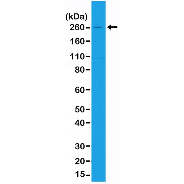
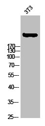
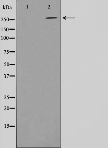
WB analysis of A431 cell lysates using GTX33608 Acetyl-CoA Carboxylase 1 antibody [RM232]. Dilution : 1:1000
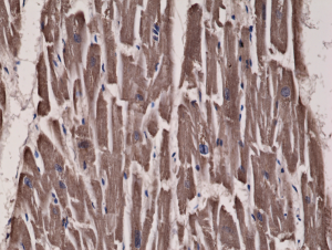
![IHC-P analysis of human heart tissue using GTX33608 Acetyl-CoA Carboxylase 1 antibody [RM232]. Dilution : 1:300](https://www.genetex.com/upload/website/prouct_img/normal/GTX33608/GTX33608_20200909_IHC-P_223_w_23060800_905.webp "IHC-P analysis of human heart tissue using GTX33608 Acetyl-CoA Carboxylase 1 antibody [RM232]. Dilution : 1:300")
WB analysis of A431 cell lysates using GTX33608 Acetyl-CoA Carboxylase 1 antibody [RM232]. Dilution : 1:1000
Acetyl-CoA Carboxylase 1 antibody [RM232]
GTX33608
ApplicationsWestern Blot, ImmunoHistoChemistry, ImmunoHistoChemistry Paraffin
Product group Antibodies
ReactivityHuman
TargetACACA
Overview
- SupplierGeneTex
- Product NameAcetyl-CoA Carboxylase 1 antibody [RM232]
- Delivery Days Customer9
- Application Supplier NoteWB: 1:1000 - 1:2000. IHC-P: 1:300 - 1:500. *Optimal dilutions/concentrations should be determined by the researcher.Not tested in other applications.
- ApplicationsWestern Blot, ImmunoHistoChemistry, ImmunoHistoChemistry Paraffin
- CertificationResearch Use Only
- ClonalityMonoclonal
- Clone IDRM232
- ConjugateUnconjugated
- Gene ID31
- Target nameACACA
- Target descriptionacetyl-CoA carboxylase alpha
- Target synonymsACAC, ACACAD, ACACalpha, ACC, ACC1, ACCA, ACCalpha, Acac1, hACC1, acetyl-CoA carboxylase 1, ACC-alpha, acetyl-Coenzyme A carboxylase alpha
- HostRabbit
- IsotypeIgG
- Protein IDQ13085
- Protein NameAcetyl-CoA carboxylase 1
- Scientific DescriptionAcetyl-CoA carboxylase (ACC) is a complex multifunctional enzyme system. ACC is a biotin-containing enzyme which catalyzes the carboxylation of acetyl-CoA to malonyl-CoA, the rate-limiting step in fatty acid synthesis. There are two ACC forms, alpha and beta, encoded by two different genes. ACC-alpha is highly enriched in lipogenic tissues. The enzyme is under long term control at the transcriptional and translational levels and under short term regulation by the phosphorylation/dephosphorylation of targeted serine residues and by allosteric transformation by citrate or palmitoyl-CoA. Multiple alternatively spliced transcript variants divergent in the 5 sequence and encoding distinct isoforms have been found for this gene. [provided by RefSeq, Jul 2008]
- ReactivityHuman
- Storage Instruction-20°C or -80°C,2°C to 8°C
- UNSPSC12352203
Datasheet
Related products
Product group Antibodies

ApplicationsWestern Blot, ELISA, ImmunoHistoChemistry
- SizePrice
Product group Antibodies
Anti-ACACA Antibody144-63833
ApplicationsImmunoFluorescence, Western Blot, ImmunoHistoChemistry
ReactivityHuman, Mouse, Rat
TargetACACA
- SizePrice
Product group Antibodies
References
ACACA Polyclonal AntibodyBS-11912R
ApplicationsImmunoFluorescence, Western Blot, ImmunoHistoChemistry, ImmunoHistoChemistry Paraffin
ReactivityBovine, Canine, Equine, Human, Mouse, Porcine, Rabbit, Rat, Sheep
- SizePrice
Product group Antibodies
Phospho-ACACA (S80) AntibodyCSB-PA009195
ApplicationsWestern Blot, ELISA, ImmunoHistoChemistry
ReactivityHuman, Mouse, Rat
TargetACACA
- SizePrice
Product group Antibodies
ApplicationsImmunoPrecipitation, Western Blot, ImmunoCytoChemistry, ImmunoHistoChemistry
TargetACACA
- SizePrice
Product group Antibodies
ApplicationsWestern Blot, ImmunoHistoChemistry
ReactivityHuman
TargetACACA
- SizePrice

Product group Antibodies
Anti-ACACA AntibodyHPA036650
ApplicationsWestern Blot, ImmunoCytoChemistry
ReactivityHuman
TargetACACA
- SizePrice
Product group Antibodies
ApplicationsWestern Blot, ImmunoHistoChemistry, ImmunoHistoChemistry Paraffin
ReactivityHuman, Mouse
TargetACACA
- SizePrice
Product group Antibodies
ApplicationsWestern Blot, ImmunoHistoChemistry
ReactivityHuman, Mouse, Rat
TargetACACA
- SizePrice