

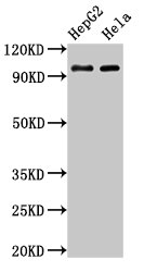
Western Blot Positive WB detected in: HepG2 whole cell lysate, Hela whole cell lysate All lanes: ACO1 antibody at 3microg/ml Secondary Goat polyclonal to rabbit IgG at 1/50000 dilution Predicted band size: 99 kDa Observed band size: 99 kDa


")
Western Blot Positive WB detected in: HepG2 whole cell lysate, Hela whole cell lysate All lanes: ACO1 antibody at 3microg/ml Secondary Goat polyclonal to rabbit IgG at 1/50000 dilution Predicted band size: 99 kDa Observed band size: 99 kDa
ACO1 Antibody
CSB-PA001161LA01HU
ApplicationsImmunoFluorescence, Western Blot, ELISA, ImmunoHistoChemistry
Product group Antibodies
ReactivityHuman
TargetACO1
Overview
- SupplierCusabio
- Product NameACO1 Antibody
- Delivery Days Customer20
- ApplicationsImmunoFluorescence, Western Blot, ELISA, ImmunoHistoChemistry
- CertificationResearch Use Only
- ClonalityPolyclonal
- ConjugateUnconjugated
- Gene ID48
- Target nameACO1
- Target descriptionaconitase 1
- Target synonymsACONS, HEL60, IREB1, IREBP, IREBP1, IRP1, cytoplasmic aconitate hydratase, aconitase 1, soluble, aconitate hydratase, cytoplasmic, citrate hydro-lyase, cytoplasmic aconitase, cytosplasmic aconitase, epididymis luminal protein 60, epididymis secretory sperm binding protein, ferritin repressor protein, iron regulatory protein 1, iron-responsive element-binding protein 1, soluble aconitase
- HostRabbit
- IsotypeIgG
- Protein IDP21399
- Protein NameCytoplasmic aconitate hydratase
- Scientific DescriptionIron sensor. Binds a 4Fe-4S cluster and functions as aconitase when cellular iron levels are high. Functions as mRNA binding protein that regulates uptake, sequestration and utilization of iron when cellular iron levels are low. Binds to iron-responsive elements (IRES) in target mRNA species when iron levels are low. Binding of a 4Fe-4S cluster precludes RNA binding.
- ReactivityHuman
- Storage Instruction-20°C or -80°C
- UNSPSC41116161
Related products
Product group Antibodies
Anti-ACO1 AntibodyA43932
ApplicationsWestern Blot
ReactivityHuman, Mouse, Rat
- SizePrice

Product group Antibodies
Anti-Aconitase 1/ACO1 Antibody Picoband(r)A02781-1-CARRIER-FREE
ApplicationsFlow Cytometry, ImmunoFluorescence, Western Blot, ELISA, ImmunoCytoChemistry, ImmunoHistoChemistry
ReactivityHuman, Monkey, Mouse, Rat
TargetACO1
- SizePrice
Product group Antibodies
Anti-ACO1 Antibody144-07867
ApplicationsWestern Blot, ImmunoHistoChemistry
ReactivityHuman, Mouse, Rat
TargetACO1
- SizePrice
Product group Antibodies
Aconitase 1 Recombinant Antibody, AbBy Fluor-647 ConjugatedBSM-62433R-BF647
ApplicationsWestern Blot
ReactivityHuman, Mouse, Rat
TargetACO1
- SizePrice
Product group Antibodies

Goat anti-ACO1 / Aconitase 1EB09487
ApplicationsWestern Blot, ELISA
ReactivityBovine, Human, Mouse, Rat
TargetACO1
- SizePrice
Product group Antibodies
Aco1 Polyclonal AntibodyCAC11187
ApplicationsImmunoFluorescence, Western Blot, ELISA, ImmunoHistoChemistry
TargetACO1
- SizePrice
Product group Antibodies

ACO1 / Aconitase AntibodyLS-C402855
ApplicationsELISA, ImmunoHistoChemistry
ReactivityHuman, Mouse, Rat
TargetACO1
- SizePrice
Product group Antibodies
Aconitase 1 antibody [N1C1]GTX102974
ApplicationsImmunoFluorescence, Western Blot, ImmunoCytoChemistry
ReactivityHuman
TargetACO1
- SizePrice

Product group Antibodies
Anti-ACO1 AntibodyHPA019371
ApplicationsWestern Blot, ImmunoCytoChemistry, ImmunoHistoChemistry
ReactivityHuman, Mouse, Rat
TargetACO1
- SizePrice