

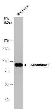
Rat tissue extract (50 μg) was separated by 7.5% SDS-PAGE, and the membrane was blotted with Aconitase 2 antibody [C1C3] (GTX109736) diluted at 1:10000.
 A: 293T B: A431 (GTX27909) 7.5% SDS PAGE GTX109736 diluted at 1:10000")
![Immunoprecipitation of Aconitase 2 protein from HeLa whole cell extracts using 5 μg of Aconitase 2 antibody [C1C3] (GTX109736). Western blot analysis was performed using Aconitase 2 antibody [C1C3] (GTX109736). EasyBlot anti-Rabbit IgG (GTX221666-01) was used as a secondary reagent.](https://www.genetex.com/upload/website/prouct_img/normal/GTX109736/GTX109736_40030_20150604_IP_w_23060500_141.webp "Immunoprecipitation of Aconitase 2 protein from HeLa whole cell extracts using 5 μg of Aconitase 2 antibody [C1C3] (GTX109736). Western blot analysis was performed using Aconitase 2 antibody [C1C3] (GTX109736). EasyBlot anti-Rabbit IgG (GTX221666-01) was used as a secondary reagent.")
![Aconitase 2 antibody [C1C3] detects Aconitase 2 protein at mitochondria by immunofluorescent analysis. Sample: HeLa cells were fixed in 4% paraformaldehyde at RT for 15 min. Green: Aconitase 2 protein stained by Aconitase 2 antibody [C1C3] (GTX109736) diluted at 1:500. Red: alpha Tubulin, a cytoskeleton marker, stained by alpha Tubulin antibody [GT114] (GTX628802) diluted at 1:500. Blue: Hoechst 33342 staining.](https://www.genetex.com/upload/website/prouct_img/normal/GTX109736/GTX109736_40030_20150410_IFA_w_23060500_645.webp "Aconitase 2 antibody [C1C3] detects Aconitase 2 protein at mitochondria by immunofluorescent analysis. Sample: HeLa cells were fixed in 4% paraformaldehyde at RT for 15 min. Green: Aconitase 2 protein stained by Aconitase 2 antibody [C1C3] (GTX109736) diluted at 1:500. Red: alpha Tubulin, a cytoskeleton marker, stained by alpha Tubulin antibody [GT114] (GTX628802) diluted at 1:500. Blue: Hoechst 33342 staining.")
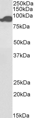
 A: mouse brain 7.5% SDS PAGE GTX109736 diluted at 1:20000")
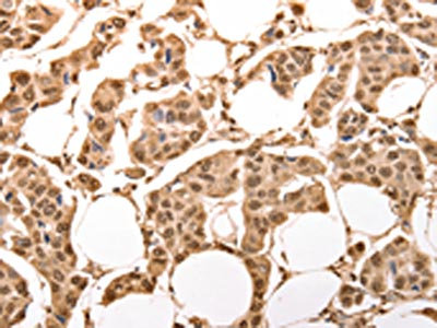
![Aconitase 2 antibody [C1C3] detects Aconitase 2 protein at mitochondria on mouse duodenum by immunohistochemical analysis. Sample: Paraffin-embedded mouse duodenum. Aconitase 2 antibody [C1C3] (GTX109736) dilution: 1:500.
Antigen Retrieval: Trilogy? (EDTA based, pH 8.0) buffer, 15min](https://www.genetex.com/upload/website/prouct_img/normal/GTX109736/GTX109736_40030_20141024_IHC_M_w_23060500_597.webp "Aconitase 2 antibody [C1C3] detects Aconitase 2 protein at mitochondria on mouse duodenum by immunohistochemical analysis. Sample: Paraffin-embedded mouse duodenum. Aconitase 2 antibody [C1C3] (GTX109736) dilution: 1:500.
Antigen Retrieval: Trilogy? (EDTA based, pH 8.0) buffer, 15min")

Rat tissue extract (50 μg) was separated by 7.5% SDS-PAGE, and the membrane was blotted with Aconitase 2 antibody [C1C3] (GTX109736) diluted at 1:10000.
Aconitase 2 antibody [C1C3]
GTX109736
ApplicationsImmunoFluorescence, ImmunoPrecipitation, Western Blot, ImmunoCytoChemistry, ImmunoHistoChemistry, ImmunoHistoChemistry Paraffin
Product group Antibodies
ReactivityHuman, Mouse, Rat
TargetACO2
Overview
- SupplierGeneTex
- Product NameAconitase 2 antibody [C1C3]
- Delivery Days Customer9
- Application Supplier NoteWB: 1:5000-1:20000. ICC/IF: 1:100-1:1000. IHC-P: 1:100-1:1000. IP: 1:100-1:500. *Optimal dilutions/concentrations should be determined by the researcher.Not tested in other applications.
- ApplicationsImmunoFluorescence, ImmunoPrecipitation, Western Blot, ImmunoCytoChemistry, ImmunoHistoChemistry, ImmunoHistoChemistry Paraffin
- CertificationResearch Use Only
- ClonalityPolyclonal
- Concentration0.82 mg/ml
- ConjugateUnconjugated
- Gene ID50
- Target nameACO2
- Target descriptionaconitase 2
- Target synonymsACONM, HEL-S-284, ICRD, OCA8, OPA9, aconitate hydratase, mitochondrial, aconitase 2, mitochondrial, citrate hydro-lyase, epididymis secretory sperm binding protein Li 284, mitochondrial aconitase
- HostRabbit
- IsotypeIgG
- Protein IDQ99798
- Protein NameAconitate hydratase, mitochondrial
- Scientific DescriptionThe protein encoded by this gene belongs to the aconitase/IPM isomerase family. It is an enzyme that catalyzes the interconversion of citrate to isocitrate via cis-aconitate in the second step of the TCA cycle. This protein is encoded in the nucleus and functions in the mitochondrion. It was found to be one of the mitochondrial matrix proteins that are preferentially degraded by the serine protease 15(PRSS15), also known as Lon protease, after oxidative modification. [provided by RefSeq]
- ReactivityHuman, Mouse, Rat
- Storage Instruction-20°C or -80°C,2°C to 8°C
- UNSPSC41116161
Datasheet
Related products
Product group Antibodies
ApplicationsWestern Blot, ELISA
ReactivityHuman
- SizePrice
Product group Antibodies
Anti-ACO2 Antibody144-03716
ApplicationsImmunoFluorescence, Western Blot
ReactivityHuman, Mouse, Rat
TargetACO2
- SizePrice
Product group Antibodies
Aconitase 2 Recombinant Antibody, AbBy Fluor-555 ConjugatedBSM-61944R-BF555
ApplicationsImmunoFluorescence, Western Blot
ReactivityHuman, Mouse, Rat
TargetACO2
- SizePrice
Product group Antibodies

Goat anti-Aconitase 2, BiotinlyatedEB09857-B
ApplicationsImmunoFluorescence, Western Blot, ELISA, ImmunoHistoChemistry
ReactivityBovine, Canine, Human, Mouse, Porcine, Rat
TargetACO2
- SizePrice
Product group Antibodies
ACO2 Polyclonal AntibodyCAC15084
ApplicationsWestern Blot, ELISA, ImmunoHistoChemistry
ReactivityMouse, Rat
TargetACO2
- SizePrice
Product group Antibodies
ACO2 AntibodyCSB-PA073168
ApplicationsWestern Blot, ELISA, ImmunoHistoChemistry
ReactivityHuman, Mouse, Rat
TargetACO2
- SizePrice
Product group Antibodies

ACO2 / Aconitase 2 AntibodyLS-C406096
ApplicationsWestern Blot, ELISA, ImmunoHistoChemistry
ReactivityHuman, Mouse, Rat
TargetACO2
- SizePrice
![WB analysis of human tissues (Lane 1-Testis ; Lane 2-Omentum ; Lane 3-Uterus ; Lane 4-Breast ; Lane 5-Brain ; Lane 6-Liver ; Lane 7-Ovary ; Lane 8-Thyroid gland ; Lane 9-colon ; Lane 10-spleen) using GTX84963 Aconitase 2 antibody [3G8]. Loading : 10 ug per lane Dilution : 1:200](https://www.genetex.com/upload/website/prouct_img/normal/GTX84963/GTX84963_4370_WB_w_23061420_802.webp)
Product group Antibodies
Aconitase 2 antibody [3G8]GTX84963
ApplicationsImmunoFluorescence, Western Blot, ImmunoCytoChemistry, ImmunoHistoChemistry, ImmunoHistoChemistry Paraffin
ReactivityHuman
TargetACO2
- SizePrice
![WB analysis of HEK293T cells transfected with Aconitase 2 plasmid (Right) or empty vector (Left) for 48 hrs using GTX84965 Aconitase 2 antibody [7G4]. Loading : 5 ug per lane](https://www.genetex.com/upload/website/prouct_img/normal/GTX84965/GTX84965_4748_WB_w_23061420_307.webp)
Product group Antibodies
Aconitase 2 antibody [7G4]GTX84965
ApplicationsFlow Cytometry, ImmunoFluorescence, Western Blot, ImmunoCytoChemistry
ReactivityHuman, Monkey, Mouse
TargetACO2
- SizePrice
![FACS analysis of Jurkat cells using GTX84966 Aconitase 2 antibody [7A11]. Red : Primary antibody Blue : Negative control antibody](https://www.genetex.com/upload/website/prouct_img/normal/GTX84966/GTX84966_644_FACS_w_23061420_395.webp)
Product group Antibodies
Aconitase 2 antibody [7A11]GTX84966
ApplicationsFlow Cytometry, ImmunoFluorescence, Western Blot, ImmunoCytoChemistry
ReactivityHuman
TargetACO2
- SizePrice