

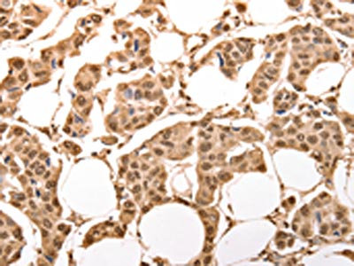
The image on the left is immunohistochemistry of paraffin-embedded Human breast cancer tissue using CSB-PA073168(ACO2 Antibody) at dilution 1/30, on the right is treated with synthetic peptide. (Original magnification: x200)
 at dilution 1/30, on the right is treated with synthetic peptide. (Original magnification: x200)")
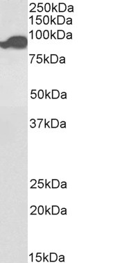
 at dilution 1/400, Secondary antibody: Goat anti rabbit IgG at 1/8000 dilution, Exposure time: 3 seconds")
The image on the left is immunohistochemistry of paraffin-embedded Human breast cancer tissue using CSB-PA073168(ACO2 Antibody) at dilution 1/30, on the right is treated with synthetic peptide. (Original magnification: x200)
ACO2 Antibody
CSB-PA073168
ApplicationsWestern Blot, ELISA, ImmunoHistoChemistry
Product group Antibodies
ReactivityHuman, Mouse, Rat
TargetACO2
Overview
- SupplierCusabio
- Product NameACO2 Antibody
- Delivery Days Customer20
- ApplicationsWestern Blot, ELISA, ImmunoHistoChemistry
- CertificationResearch Use Only
- ClonalityPolyclonal
- ConjugateUnconjugated
- Gene ID50
- Target nameACO2
- Target descriptionaconitase 2
- Target synonymsACONM, HEL-S-284, ICRD, OCA8, OPA9, aconitate hydratase, mitochondrial, aconitase 2, mitochondrial, citrate hydro-lyase, epididymis secretory sperm binding protein Li 284, mitochondrial aconitase
- HostRabbit
- IsotypeIgG
- Protein IDQ99798
- Protein NameAconitate hydratase, mitochondrial
- Scientific DescriptionThe protein encoded by this gene belongs to the aconitase/IPM isomerase family. It is an enzyme that catalyzes the interconversion of citrate to isocitrate via cis-aconitate in the second step of the TCA cycle. This protein is encoded in the nucleus and functions in the mitochondrion. It was found to be one of the mitochondrial matrix proteins that are preferentially degraded by the serine protease 15(PRSS15), also known as Lon protease, after oxidative modification.
- ReactivityHuman, Mouse, Rat
- Storage Instruction-20°C or -80°C
- UNSPSC41116161
Related products
Product group Antibodies
ApplicationsWestern Blot, ELISA
ReactivityHuman
- SizePrice
Product group Antibodies
Anti-ACO2 Antibody144-03716
ApplicationsImmunoFluorescence, Western Blot
ReactivityHuman, Mouse, Rat
TargetACO2
- SizePrice
Product group Antibodies
Aconitase 2 Recombinant Antibody, AbBy Fluor-555 ConjugatedBSM-61944R-BF555
ApplicationsImmunoFluorescence, Western Blot
ReactivityHuman, Mouse, Rat
TargetACO2
- SizePrice
Product group Antibodies

Goat anti-Aconitase 2, BiotinlyatedEB09857-B
ApplicationsImmunoFluorescence, Western Blot, ELISA, ImmunoHistoChemistry
ReactivityBovine, Canine, Human, Mouse, Porcine, Rat
TargetACO2
- SizePrice
Product group Antibodies
ACO2 Polyclonal AntibodyCAC15084
ApplicationsWestern Blot, ELISA, ImmunoHistoChemistry
ReactivityMouse, Rat
TargetACO2
- SizePrice
Product group Antibodies

ACO2 / Aconitase 2 AntibodyLS-C406096
ApplicationsWestern Blot, ELISA, ImmunoHistoChemistry
ReactivityHuman, Mouse, Rat
TargetACO2
- SizePrice

Product group Antibodies
Anti-ACO2 AntibodyHPA001097
ApplicationsWestern Blot, ImmunoCytoChemistry, ImmunoHistoChemistry
ReactivityHuman, Mouse, Rat
TargetACO2
- SizePrice
![Rat tissue extract (50 μg) was separated by 7.5% SDS-PAGE, and the membrane was blotted with Aconitase 2 antibody [C1C3] (GTX109736) diluted at 1:10000.](https://www.genetex.com/upload/website/prouct_img/normal/GTX109736/GTX109736_40030_20160310_WB_R_brain_w_23060500_720.webp)
Product group Antibodies
Aconitase 2 antibody [C1C3]GTX109736
ApplicationsImmunoFluorescence, ImmunoPrecipitation, Western Blot, ImmunoCytoChemistry, ImmunoHistoChemistry, ImmunoHistoChemistry Paraffin
ReactivityHuman, Mouse, Rat
TargetACO2
- SizePrice
Product group Antibodies
ApplicationsWestern Blot, ELISA, ImmunoHistoChemistry, ImmunoHistoChemistry Paraffin
ReactivityHuman
TargetACO2
- SizePrice