

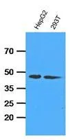
WB analysis of indicated lysates (35ug per lane) using ADK antibody at a dilution of 1:1,000.
WB analysis of indicated lysates (35ug per lane) using ADK antibody at a dilution of 1:1,000.
Adenosine kinase antibody [AT4F8]
GTX53683
ApplicationsWestern Blot, ELISA
Product group Antibodies
ReactivityHuman
TargetADK
Overview
- SupplierGeneTex
- Product NameAdenosine kinase antibody [AT4F8]
- Delivery Days Customer9
- Application Supplier NoteThe antibody has been tested by ELISA and Western blot analysis to assure specificity and reactivity. Since application varies, however, each investigation should be titrated by the reagent to obtain optimal results. Recommended dilution range for Western blot analysis is 1:1000. Recommended starting dilution is 1:1000
- ApplicationsWestern Blot, ELISA
- CertificationResearch Use Only
- ClonalityMonoclonal
- Clone IDAT4F8
- Concentration1 mg/ml
- ConjugateUnconjugated
- Gene ID132
- Target nameADK
- Target descriptionadenosine kinase
- Target synonymsAK, adenosine kinase, adenosine 5'-phosphotransferase, testicular tissue protein Li 14
- HostMouse
- IsotypeIgG1
- Protein IDP55263
- Protein NameAdenosine kinase
- Scientific DescriptionThis gene an enzyme which catalyzes the transfer of the gamma-phosphate from ATP to adenosine, thereby serving as a regulator of concentrations of both extracellular adenosine and intracellular adenine nucleotides. Adenosine has widespread effects on the cardiovascular, nervous, respiratory, and immune systems and inhibitors of the enzyme could play an important pharmacological role in increasing intravascular adenosine concentrations and acting as anti-inflammatory agents. Multiple transcript variants encoding different isoforms have been found for this gene. [provided by RefSeq, Jan 2011]
- ReactivityHuman
- Storage Instruction-20°C or -80°C,2°C to 8°C
- UNSPSC41116161
Datasheet
Related products
Product group Antibodies
Anti-ADK AntibodyA98547
ApplicationsWestern Blot, ELISA
ReactivityHuman, Mouse, Rat
- SizePrice
Product group Antibodies

ADK / Adenosine Kinase AntibodyLS-C749996
ApplicationsWestern Blot
ReactivityHuman, Mouse, Rat
TargetADK
- SizePrice
Product group Antibodies
ADK Recombinant AntibodyBSM-60657R
ApplicationsImmunoFluorescence, Western Blot, ImmunoHistoChemistry, ImmunoHistoChemistry Frozen, ImmunoHistoChemistry Paraffin
ReactivityHuman, Mouse, Rat
TargetADK
- SizePrice
Product group Antibodies
ADK AntibodyCSB-PA152141
ApplicationsWestern Blot, ELISA
ReactivityHuman, Mouse, Rat
TargetADK
- SizePrice
Product group Antibodies
Adk Polyclonal AntibodyCAC11718
ApplicationsImmunoFluorescence, Western Blot, ELISA, ImmunoHistoChemistry
ReactivityMouse, Rat
TargetADK
- SizePrice
![Wild-type (WT) and Adenosine kinase knockout (KO) HeLa cell extracts (30 μg) were separated by 10% SDS-PAGE, and the membrane was blotted with Adenosine kinase antibody [N1C1] (GTX101372) diluted at 1:500. The HRP-conjugated anti-rabbit IgG antibody (GTX213110-01) was used to detect the primary antibody.](https://www.genetex.com/upload/website/prouct_img/normal/GTX101372/GTX101372_40583_20180810_WB_KO_watermark_w_23060100_591.webp)
Product group Antibodies
Adenosine kinase antibody [N1C1]GTX101372
ApplicationsImmunoFluorescence, Western Blot, ImmunoCytoChemistry
ReactivityHuman
TargetADK
- SizePrice
Product group Antibodies
Adenosine kinase antibodyGTX101385
ApplicationsImmunoFluorescence, Western Blot, ImmunoCytoChemistry, ImmunoHistoChemistry, ImmunoHistoChemistry Paraffin
ReactivityHuman, Mouse, Rat
TargetADK
- SizePrice

Product group Antibodies
Anti-ADK AntibodyHPA038391
ApplicationsImmunoHistoChemistry
ReactivityHuman
TargetADK
- SizePrice