

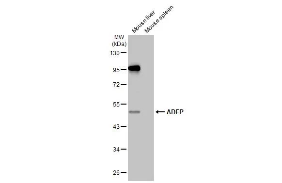
Various tissue extracts (50 μg) were separated by 10% SDS-PAGE, and the membrane was blotted with ADFP antibody [HL2147] (GTX638125) diluted at 1:1000. The HRP-conjugated anti-rabbit IgG antibody (GTX213110-01) was used to detect the primary antibody.
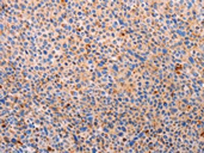
![ADFP antibody [HL2147] detects ADFP protein by immunohistochemical analysis. Sample: Paraffin-embedded mouse tissues. ADFP stained by ADFP antibody [HL2147] (GTX638125) diluted at 1:100. Antigen Retrieval: Citrate buffer, pH 6.0, 15 min](https://www.genetex.com/upload/website/prouct_img/normal/GTX638125/GTX638125_T-44928_20230217_IHC-P_multiple_M_23021401_288.webp "ADFP antibody [HL2147] detects ADFP protein by immunohistochemical analysis. Sample: Paraffin-embedded mouse tissues. ADFP stained by ADFP antibody [HL2147] (GTX638125) diluted at 1:100. Antigen Retrieval: Citrate buffer, pH 6.0, 15 min")
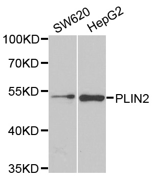
![Various whole cell extracts (30 μg) were separated by 10% SDS-PAGE, and the membrane was blotted with ADFP antibody [HL2147] (GTX638125) diluted at 1:1000. The HRP-conjugated anti-rabbit IgG antibody (GTX213110-01) was used to detect the primary antibody.](https://www.genetex.com/upload/website/prouct_img/normal/GTX638125/GTX638125_45012_20230421_WB_23042500_668.webp "Various whole cell extracts (30 μg) were separated by 10% SDS-PAGE, and the membrane was blotted with ADFP antibody [HL2147] (GTX638125) diluted at 1:1000. The HRP-conjugated anti-rabbit IgG antibody (GTX213110-01) was used to detect the primary antibody.")
![Whole cell extract (30 μg) was separated by 10% SDS-PAGE, and the membrane was blotted with ADFP antibody [HL2147] (GTX638125) diluted at 1:1000. The HRP-conjugated anti-rabbit IgG antibody (GTX213110-01) was used to detect the primary antibody.](https://www.genetex.com/upload/website/prouct_img/normal/GTX638125/GTX638125_45012_20230526_WB_R_23053001_848.webp "Whole cell extract (30 μg) was separated by 10% SDS-PAGE, and the membrane was blotted with ADFP antibody [HL2147] (GTX638125) diluted at 1:1000. The HRP-conjugated anti-rabbit IgG antibody (GTX213110-01) was used to detect the primary antibody.")
![ADFP antibody [HL2147] detects ADFP protein at lipid droplet by immunofluorescent analysis. Sample: Mock and differentiated 3T3-L1 cells were fixed in 4% paraformaldehyde at RT for 15 min. Green: ADFP stained by ADFP antibody [HL2147] (GTX638125) diluted at 1:500. Blue: Fluoroshield with DAPI (GTX30920).](https://www.genetex.com/upload/website/prouct_img/normal/GTX638125/GTX638125_45012_20231006_ICC_IF_M_differentiation_23102401_900.webp "ADFP antibody [HL2147] detects ADFP protein at lipid droplet by immunofluorescent analysis. Sample: Mock and differentiated 3T3-L1 cells were fixed in 4% paraformaldehyde at RT for 15 min. Green: ADFP stained by ADFP antibody [HL2147] (GTX638125) diluted at 1:500. Blue: Fluoroshield with DAPI (GTX30920).")
Various tissue extracts (50 μg) were separated by 10% SDS-PAGE, and the membrane was blotted with ADFP antibody [HL2147] (GTX638125) diluted at 1:1000. The HRP-conjugated anti-rabbit IgG antibody (GTX213110-01) was used to detect the primary antibody.
ADFP antibody [HL2147]
GTX638125
ApplicationsImmunoFluorescence, Western Blot, ImmunoCytoChemistry, ImmunoHistoChemistry, ImmunoHistoChemistry Paraffin
Product group Antibodies
ReactivityHuman, Mouse, Rat
TargetPLIN2
Overview
- SupplierGeneTex
- Product NameADFP antibody [HL2147]
- Delivery Days Customer9
- ApplicationsImmunoFluorescence, Western Blot, ImmunoCytoChemistry, ImmunoHistoChemistry, ImmunoHistoChemistry Paraffin
- CertificationResearch Use Only
- ClonalityMonoclonal
- Clone IDHL2147
- Concentration1 mg/ml
- ConjugateUnconjugated
- Gene ID123
- Target namePLIN2
- Target descriptionperilipin 2
- Target synonymsADFP, ADRP, perilipin-2, adipophilin, adipose differentiation-related protein
- HostRabbit
- IsotypeIgG
- Protein IDQ99541
- Protein NamePerilipin-2
- Scientific DescriptionThe protein encoded by this gene belongs to the perilipin family, members of which coat intracellular lipid storage droplets. This protein is associated with the lipid globule surface membrane material, and maybe involved in development and maintenance of adipose tissue. However, it is not restricted to adipocytes as previously thought, but is found in a wide range of cultured cell lines, including fibroblasts, endothelial and epithelial cells, and tissues, such as lactating mammary gland, adrenal cortex, Sertoli and Leydig cells, and hepatocytes in alcoholic liver cirrhosis, suggesting that it may serve as a marker of lipid accumulation in diverse cell types and diseases. Alternatively spliced transcript variants have been found for this gene. [provided by RefSeq, Mar 2011]
- ReactivityHuman, Mouse, Rat
- Storage Instruction-20°C or -80°C,2°C to 8°C
- UNSPSC41116161
Datasheet
Related products
Product group Antibodies
PLIN2 AntibodyCSB-PA065885
ApplicationsWestern Blot, ELISA, ImmunoHistoChemistry
ReactivityHuman, Mouse
TargetPLIN2
- SizePrice
Product group Antibodies
ApplicationsImmunoPrecipitation, Western Blot, ImmunoCytoChemistry, ImmunoHistoChemistry
TargetPLIN2
- SizePrice
Product group Antibodies
Anti-PLIN2 Antibody144-06276
ApplicationsWestern Blot
ReactivityHuman, Mouse, Rat
TargetPLIN2
- SizePrice

Product group Antibodies
Anti-PLIN2 AntibodyHPA016607
ApplicationsWestern Blot, ImmunoCytoChemistry, ImmunoHistoChemistry
ReactivityHuman
TargetPLIN2
- SizePrice
Product group Antibodies

PLIN2 / ADFP / Adipophilin AntibodyLS-C402989
ApplicationsWestern Blot, ELISA, ImmunoHistoChemistry
ReactivityHuman, Mouse
TargetPLIN2
- SizePrice

Product group Antibodies
Anti-ADFP/PLIN2 Antibody Picoband(r)PB10083-CARRIER-FREE
ApplicationsWestern Blot, ImmunoHistoChemistry
ReactivityHuman, Mouse, Rat
TargetPLIN2
- SizePrice
![Various tissue extracts (50 μg) were separated by 10% SDS-PAGE, and the membrane was blotted with ADFP antibody [HL2145] (GTX638123) diluted at 1:1000. The HRP-conjugated anti-rabbit IgG antibody (GTX213110-01) was used to detect the primary antibody.](https://www.genetex.com/upload/website/prouct_img/normal/GTX638123/GTX638123_T-44928_20230120_WB_M_tissue_23013122_704.webp)
Product group Antibodies
ADFP antibody [HL2145]GTX638123
ApplicationsWestern Blot, ImmunoHistoChemistry, ImmunoHistoChemistry Paraffin
ReactivityHuman, Mouse
TargetPLIN2
- SizePrice