

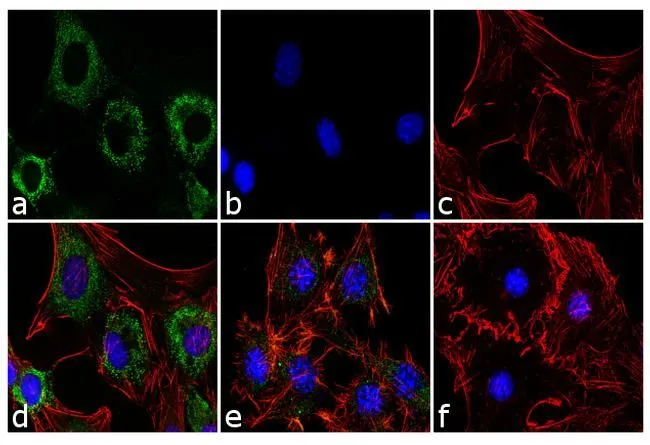
ICC/IF analysis of differentiated 3T3-L1 cells (treated with StemPro Adipogenesis Supplement for 5 days) using GTX80683 Adiponectin antibody [19F1]. Panel e is untreated cell with less signal. Panel f represents control cells with no primary antibody to assess background. Green : Primary antibody Blue : Nuclei Red : Actin Fixation : 4% paraformaldehyde Permeabilization : 0.1% Triton X-100 for 10 minute Dilution : 2 μg/ml in 0.1% BSA incubated for 3 hours at room temperature
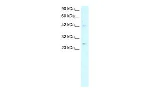
![WB analysis of rabbit subcutaneous white adipose tissue (scWAT), gonadal white adipose tissue (gWAT), perirenal white adipose tissue (pWAT), tibia marrow adipose tissue (tibMAT), tibia red marrow (tibRM), and 3T3-L1 positive control adipocyte lysates using GTX80683 Adiponectin antibody [19F1]. Dilution : 1:1000](https://www.genetex.com/upload/website/prouct_img/normal/GTX80683/GTX80683_1948_WB_w_23061322_328.webp "WB analysis of rabbit subcutaneous white adipose tissue (scWAT), gonadal white adipose tissue (gWAT), perirenal white adipose tissue (pWAT), tibia marrow adipose tissue (tibMAT), tibia red marrow (tibRM), and 3T3-L1 positive control adipocyte lysates using GTX80683 Adiponectin antibody [19F1]. Dilution : 1:1000")
![IHC-P analysis of human skin tissue using GTX80683 Adiponectin antibody [19F1]. Left : Primary antibody Right : Negative control without primary antibody Antigen retrieval : heat induced antigen retrieval was performed using 10mM sodium citrate (pH6.0) buffer, microwaved for 8-15 minutes Dilution : 1:20](https://www.genetex.com/upload/website/prouct_img/normal/GTX80683/GTX80683_1406_IHC-P_w_23061322_599.webp "IHC-P analysis of human skin tissue using GTX80683 Adiponectin antibody [19F1]. Left : Primary antibody Right : Negative control without primary antibody Antigen retrieval : heat induced antigen retrieval was performed using 10mM sodium citrate (pH6.0) buffer, microwaved for 8-15 minutes Dilution : 1:20")
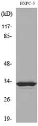
![WB analysis of 10 μl of conditioned media of HepG2 cell using GTX80683 Adiponectin antibody [19F1]. Dilution : 2 μg/ml](https://www.genetex.com/upload/website/prouct_img/normal/GTX80683/GTX80683_1950_WB_w_23061322_621.webp "WB analysis of 10 μl of conditioned media of HepG2 cell using GTX80683 Adiponectin antibody [19F1]. Dilution : 2 μg/ml")
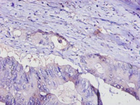
![IHC-P analysis of human colon carcinoma tissue using GTX80683 Adiponectin antibody [19F1]. Left : Primary antibody Right : Negative control without primary antibody Antigen retrieval : heat induced antigen retrieval was performed using 10mM sodium citrate (pH6.0) buffer, microwaved for 8-15 minutes Dilution : 1:100](https://www.genetex.com/upload/website/prouct_img/normal/GTX80683/GTX80683_1407_IHC-P_w_23061322_267.webp "IHC-P analysis of human colon carcinoma tissue using GTX80683 Adiponectin antibody [19F1]. Left : Primary antibody Right : Negative control without primary antibody Antigen retrieval : heat induced antigen retrieval was performed using 10mM sodium citrate (pH6.0) buffer, microwaved for 8-15 minutes Dilution : 1:100")
ICC/IF analysis of differentiated 3T3-L1 cells (treated with StemPro Adipogenesis Supplement for 5 days) using GTX80683 Adiponectin antibody [19F1]. Panel e is untreated cell with less signal. Panel f represents control cells with no primary antibody to assess background. Green : Primary antibody Blue : Nuclei Red : Actin Fixation : 4% paraformaldehyde Permeabilization : 0.1% Triton X-100 for 10 minute Dilution : 2 μg/ml in 0.1% BSA incubated for 3 hours at room temperature
Adiponectin antibody [19F1]
GTX80683
ApplicationsImmunoFluorescence, Western Blot, ELISA, ImmunoCytoChemistry, ImmunoHistoChemistry, ImmunoHistoChemistry Paraffin
Product group Antibodies
ReactivityHuman, Mouse, Rabbit
TargetADIPOQ
Overview
- SupplierGeneTex
- Product NameAdiponectin antibody [19F1]
- Delivery Days Customer9
- Application Supplier NoteWB: 2 microg/ml. ICC/IF: 2-4 microg/ml. *Optimal dilutions/concentrations should be determined by the researcher.Not tested in other applications.
- ApplicationsImmunoFluorescence, Western Blot, ELISA, ImmunoCytoChemistry, ImmunoHistoChemistry, ImmunoHistoChemistry Paraffin
- CertificationResearch Use Only
- ClonalityMonoclonal
- Clone ID19F1
- Concentration1 mg/ml
- ConjugateUnconjugated
- Gene ID9370
- Target nameADIPOQ
- Target descriptionadiponectin, C1Q and collagen domain containing
- Target synonymsACDC, ACRP30, ADIPQTL1, ADPN, APM-1, APM1, GBP28, adiponectin, 30 kDa adipocyte complement-related protein, adipocyte complement-related 30 kDa protein, adipose most abundant gene transcript 1 protein, adipose specific collagen-like factor, gelatin-binding protein 28
- HostMouse
- IsotypeIgG
- Protein IDQ15848
- Protein NameAdiponectin
- Scientific DescriptionThis gene is expressed in adipose tissue exclusively. It encodes a protein with similarity to collagens X and VIII and complement factor C1q. The encoded protein circulates in the plasma and is involved with metabolic and hormonal processes. Mutations in this gene are associated with adiponectin deficiency. Multiple alternatively spliced variants, encoding the same protein, have been identified. [provided by RefSeq, Apr 2010]
- ReactivityHuman, Mouse, Rabbit
- Storage Instruction-20°C or -80°C,2°C to 8°C
- UNSPSC41116161
References
- A mouse model of human mitofusin-2-related lipodystrophy exhibits adipose-specific mitochondrial stress and reduced leptin secretion.Read this paper
- Essential Amino Acids-Rich Diet Decreased Adipose Tissue Storage in Adult Mice: A Preliminary Histopathological Study.Read this paper
Datasheet
Related products
Product group Antibodies
ADIPOQ AntibodyCSB-PA07956A0RB
ApplicationsWestern Blot, ELISA, ImmunoHistoChemistry
ReactivityHuman, Mouse
TargetADIPOQ
- SizePrice
Product group Antibodies
Adipoq Polyclonal AntibodyCAC10447
ApplicationsWestern Blot, ELISA, ImmunoHistoChemistry
ReactivityMouse
TargetADIPOQ
- SizePrice
Product group Antibodies
Anti-F3 AntibodyA101658
ApplicationsWestern Blot, ELISA
ReactivityHuman
- SizePrice
Product group Antibodies
Anti-Acrp30 Antibody130-10074
ApplicationsELISA
ReactivityHuman
TargetADIPOQ
- SizePrice
Product group Antibodies

ACDC AntibodyABX122695
ApplicationsWestern Blot, ELISA, ImmunoHistoChemistry
- SizePrice
Product group Antibodies

Adiponectin Antibody (Preservative Free)LS-C743763
ApplicationsWestern Blot, ELISA
ReactivityHuman
TargetADIPOQ
- SizePrice

Product group Antibodies
Anti-ADIPOQ AntibodyHPA051767
ApplicationsWestern Blot, ImmunoHistoChemistry
ReactivityHuman
TargetADIPOQ
- SizePrice

Product group Antibodies
Anti-Adiponectin/ADIPOQ Antibody Picoband(r)PB9001-CARRIER-FREE
ApplicationsWestern Blot, ImmunoHistoChemistry
ReactivityHuman
TargetADIPOQ
- SizePrice
Product group Antibodies
Adiponectin antibody, N-termGTX77828
ApplicationsWestern Blot, ImmunoHistoChemistry, ImmunoHistoChemistry Paraffin
ReactivityHuman
TargetADIPOQ
- SizePrice