

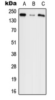
WB analysis of HEK293T (A), NIH3T3 (B), PC12 (C) whole cell lysates using GTX54904 Agrin antibody.
WB analysis of HEK293T (A), NIH3T3 (B), PC12 (C) whole cell lysates using GTX54904 Agrin antibody.
Agrin antibody
GTX54904
ApplicationsWestern Blot, ImmunoHistoChemistry, ImmunoHistoChemistry Paraffin
Product group Antibodies
ReactivityHuman, Mouse, Rat
TargetAGRN
Overview
- SupplierGeneTex
- Product NameAgrin antibody
- Delivery Days Customer9
- Application Supplier NoteWB: 1:500 - 1:1000. *Optimal dilutions/concentrations should be determined by the researcher.Not tested in other applications.
- ApplicationsWestern Blot, ImmunoHistoChemistry, ImmunoHistoChemistry Paraffin
- CertificationResearch Use Only
- ClonalityPolyclonal
- ConjugateUnconjugated
- Gene ID375790
- Target nameAGRN
- Target descriptionagrin
- Target synonymsAGRIN, CMS8, CMSPPD, agrin, agrin proteoglycan
- HostRabbit
- IsotypeIgG
- Protein IDO00468
- Protein NameAgrin
- Scientific DescriptionThis gene encodes one of several proteins that are critical in the development of the neuromuscular junction (NMJ), as identified in mouse knock-out studies. The encoded protein contains several laminin G, Kazal type serine protease inhibitor, and epidermal growth factor domains. Additional post-translational modifications occur to add glycosaminoglycans and disulfide bonds. In one family with congenital myasthenic syndrome affecting limb-girdle muscles, a mutation in this gene was found. Alternative splicing results in multiple transcript variants encoding different isoforms. [provided by RefSeq, Mar 2015]
- ReactivityHuman, Mouse, Rat
- Storage Instruction-20°C or -80°C,2°C to 8°C
- UNSPSC41116161
References
- Determination of Agrin and Related Proteins Levels as a Function of Age in Human Hearts. Skeffington KL et al., 2022, Front Cardiovasc MedRead this paper
- Mass-spectrometry analysis of the human pineal proteome during night and day and in autism. Dumas G et al., 2021 Apr, J Pineal ResRead this paper
Datasheet
Related products
Product group Antibodies
Anti-AGRN AntibodyA28287
ApplicationsWestern Blot, ImmunoHistoChemistry
ReactivityHuman, Mouse, Rat
- SizePrice

Product group Antibodies
Anti-Agrin/AGRN Picoband(r) AntibodyA04649-CARRIER-FREE
ApplicationsFlow Cytometry, ImmunoFluorescence, Western Blot, ELISA, ImmunoCytoChemistry, ImmunoHistoChemistry
ReactivityHuman, Mouse, Rat
TargetAGRN
- SizePrice
Product group Antibodies
Anti-AGRN Antibody144-64880
ApplicationsWestern Blot
ReactivityHuman, Mouse, Rat
TargetAGRN
- SizePrice
Product group Antibodies
AGRN AntibodyCSB-PA001461LA01HU
ApplicationsImmunoFluorescence, Western Blot, ELISA, ImmunoHistoChemistry
ReactivityHuman
TargetAGRN
- SizePrice
Product group Antibodies
Agrn Polyclonal AntibodyCAC11305
ApplicationsImmunoFluorescence, Western Blot, ELISA, ImmunoHistoChemistry
TargetAGRN
- SizePrice

Product group Antibodies
Anti-AGRN AntibodyHPA040090
ApplicationsImmunoCytoChemistry, ImmunoHistoChemistry
ReactivityHuman
TargetAGRN
- SizePrice
Product group Antibodies
Anti-AGRNY058325
ApplicationsELISA, ImmunoHistoChemistry
ReactivityHuman, Mouse, Rat
- SizePrice
Product group Antibodies

AGRN / Agrin AntibodyLS-C402607
ApplicationsELISA, ImmunoHistoChemistry
ReactivityHuman
TargetAGRN
- SizePrice