

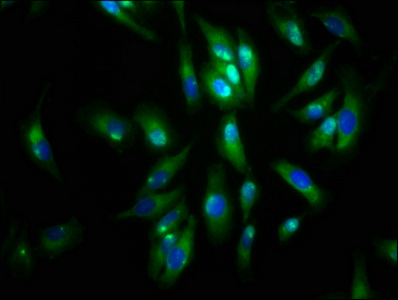
Immunofluorescence staining of Hela cells with CSB-PA001461LA01HU at 1:133, counter-stained with DAPI. The cells were fixed in 4% formaldehyde, permeabilized using 0.2% Triton X-100 and blocked in 10% normal Goat Serum. The cells were then incubated with the antibody overnight at 4°C. The secondary antibody was Alexa Fluor 488-congugated AffiniPure Goat Anti-Rabbit IgG(H+L).

. Section was blocked with 10% normal goat serum 30min at RT. Then primary antibody (1% BSA) was incubated at 4°C overnight. The primary is detected by a biotinylated secondary antibody and visualized using an HRP conjugated SP system.")
. Section was blocked with 10% normal goat serum 30min at RT. Then primary antibody (1% BSA) was incubated at 4°C overnight. The primary is detected by a biotinylated secondary antibody and visualized using an HRP conjugated SP system.")
Immunofluorescence staining of Hela cells with CSB-PA001461LA01HU at 1:133, counter-stained with DAPI. The cells were fixed in 4% formaldehyde, permeabilized using 0.2% Triton X-100 and blocked in 10% normal Goat Serum. The cells were then incubated with the antibody overnight at 4°C. The secondary antibody was Alexa Fluor 488-congugated AffiniPure Goat Anti-Rabbit IgG(H+L).
AGRN Antibody
CSB-PA001461LA01HU
ApplicationsImmunoFluorescence, Western Blot, ELISA, ImmunoHistoChemistry
Product group Antibodies
ReactivityHuman
TargetAGRN
Overview
- SupplierCusabio
- Product NameAGRN Antibody
- Delivery Days Customer20
- ApplicationsImmunoFluorescence, Western Blot, ELISA, ImmunoHistoChemistry
- CertificationResearch Use Only
- ClonalityPolyclonal
- ConjugateUnconjugated
- Gene ID375790
- Target nameAGRN
- Target descriptionagrin
- Target synonymsAGRIN, CMS8, CMSPPD, agrin, agrin proteoglycan
- HostRabbit
- IsotypeIgG
- Protein IDO00468
- Protein NameAgrin
- Scientific DescriptionIsoform 1: heparan sulfate basal lamina glycoprotein that plays a central role in the formation and the maintenance of the neuromuscular junction (NMJ) and directs key events in postsynaptic differentiation. Component of the AGRN-LRP4 receptor complex that induces the phosphorylation and activation of MUSK. The activation of MUSK in myotubes induces the formation of NMJ by regulating different processes including the transcription of specific genes and the clustering of AChR in the postsynaptic membrane. Calcium ions are required for maximal AChR clustering. AGRN function in neurons is highly regulated by alternative splicing, glycan binding and proteolytic processing. Modulates calcium ion homeostasis in neurons, specifically by inducing an increase in cytoplasmic calcium ions. Functions differentially in the central nervous system (CNS) by inhibiting the alpha(3)-subtype of Na+/K+-ATPase and evoking depolarization at CNS synapses. This secreted isoform forms a bridge, after release from motor neurons, to basal lamina through binding laminin via the NtA domain.
- ReactivityHuman
- Storage Instruction-20°C or -80°C
- UNSPSC41116161
Related products
Product group Antibodies
Anti-AGRN AntibodyA28287
ApplicationsWestern Blot, ImmunoHistoChemistry
ReactivityHuman, Mouse, Rat
- SizePrice

Product group Antibodies
Anti-Agrin/AGRN Picoband(r) AntibodyA04649-CARRIER-FREE
ApplicationsFlow Cytometry, ImmunoFluorescence, Western Blot, ELISA, ImmunoCytoChemistry, ImmunoHistoChemistry
ReactivityHuman, Mouse, Rat
TargetAGRN
- SizePrice
Product group Antibodies
Anti-AGRN Antibody144-64880
ApplicationsWestern Blot
ReactivityHuman, Mouse, Rat
TargetAGRN
- SizePrice
Product group Antibodies
Agrn Polyclonal AntibodyCAC11305
ApplicationsImmunoFluorescence, Western Blot, ELISA, ImmunoHistoChemistry
TargetAGRN
- SizePrice

Product group Antibodies
Anti-AGRN AntibodyHPA040090
ApplicationsImmunoCytoChemistry, ImmunoHistoChemistry
ReactivityHuman
TargetAGRN
- SizePrice
Product group Antibodies
References
Agrin antibodyGTX54904
ApplicationsWestern Blot, ImmunoHistoChemistry, ImmunoHistoChemistry Paraffin
ReactivityHuman, Mouse, Rat
TargetAGRN
- SizePrice
Product group Antibodies
Anti-AGRNY058325
ApplicationsELISA, ImmunoHistoChemistry
ReactivityHuman, Mouse, Rat
- SizePrice
Product group Antibodies

AGRN / Agrin AntibodyLS-C402607
ApplicationsELISA, ImmunoHistoChemistry
ReactivityHuman
TargetAGRN
- SizePrice