

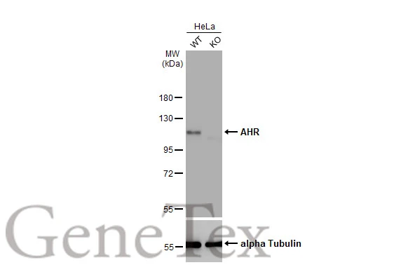
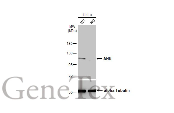
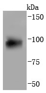
Wild-type (WT) and AHR knockout (KO) HeLa cell extracts (30 μg) were separated by 7.5% SDS-PAGE, and the membrane was blotted with AHR antibody [HL1984] (GTX637885) diluted at 1:1000. The HRP-conjugated anti-rabbit IgG antibody (GTX213110-01) was used to detect the primary antibody.
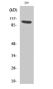
![Various whole cell extracts (30 μg) were separated by 5% SDS-PAGE, and the membrane was blotted with AHR antibody [HL1984] (GTX637885) diluted at 1:500. The HRP-conjugated anti-rabbit IgG antibody (GTX213110-01) was used to detect the primary antibody.](https://www.genetex.com/upload/website/prouct_img/normal/GTX637885/GTX637885_T-44872_20221223_WB_M_22122722_403.webp "Various whole cell extracts (30 μg) were separated by 5% SDS-PAGE, and the membrane was blotted with AHR antibody [HL1984] (GTX637885) diluted at 1:500. The HRP-conjugated anti-rabbit IgG antibody (GTX213110-01) was used to detect the primary antibody.")
![AHR antibody [HL1984] detects AHR protein at cytoplasm and nucleus by immunofluorescent analysis. Sample: U87-MG cells were fixed in 4% paraformaldehyde at RT for 15 min. Green: AHR stained by AHR antibody [HL1984] (GTX637885) diluted at 1:500. Blue: Fluoroshield with DAPI (GTX30920).](https://www.genetex.com/upload/website/prouct_img/normal/GTX637885/GTX637885_T-44872_20230203_ICC_IF_23021401_172.webp "AHR antibody [HL1984] detects AHR protein at cytoplasm and nucleus by immunofluorescent analysis. Sample: U87-MG cells were fixed in 4% paraformaldehyde at RT for 15 min. Green: AHR stained by AHR antibody [HL1984] (GTX637885) diluted at 1:500. Blue: Fluoroshield with DAPI (GTX30920).")
![AHR antibody [HL1984] detects AHR protein at cytoplasm and nucleus by immunofluorescent analysis. Sample: HeLa cells were fixed in 4% paraformaldehyde at RT for 15 min. Green: AHR stained by AHR antibody [HL1984] (GTX637885) diluted at 1:500. Blue: Fluoroshield with DAPI (GTX30920).](https://www.genetex.com/upload/website/prouct_img/normal/GTX637885/GTX637885_T-44872_20230331_ICC_IF_23041023_715.webp "AHR antibody [HL1984] detects AHR protein at cytoplasm and nucleus by immunofluorescent analysis. Sample: HeLa cells were fixed in 4% paraformaldehyde at RT for 15 min. Green: AHR stained by AHR antibody [HL1984] (GTX637885) diluted at 1:500. Blue: Fluoroshield with DAPI (GTX30920).")
![Various whole cell extracts (30 μg) were separated by 5% SDS-PAGE, and the membrane was blotted with AHR antibody [HL1984] (GTX637885) diluted at 1:1000. The HRP-conjugated anti-rabbit IgG antibody (GTX213110-01) was used to detect the primary antibody. Corresponding RNA expression data for the same cell lines are based on Human Protein Atlas program.](https://www.genetex.com/upload/website/prouct_img/normal/GTX637885/GTX637885_45278_20240119_WB_TPM_watermark_24112622_378.webp "Various whole cell extracts (30 μg) were separated by 5% SDS-PAGE, and the membrane was blotted with AHR antibody [HL1984] (GTX637885) diluted at 1:1000. The HRP-conjugated anti-rabbit IgG antibody (GTX213110-01) was used to detect the primary antibody. Corresponding RNA expression data for the same cell lines are based on Human Protein Atlas program.")
Wild-type (WT) and AHR knockout (KO) HeLa cell extracts (30 μg) were separated by 7.5% SDS-PAGE, and the membrane was blotted with AHR antibody [HL1984] (GTX637885) diluted at 1:1000. The HRP-conjugated anti-rabbit IgG antibody (GTX213110-01) was used to detect the primary antibody.
AHR antibody [HL1984]
GTX637885
ApplicationsImmunoFluorescence, Western Blot, ImmunoCytoChemistry
Product group Antibodies
ReactivityHuman, Mouse
TargetAHR
Overview
- SupplierGeneTex
- Product NameAHR antibody [HL1984]
- Delivery Days Customer9
- Application Supplier NoteWB: 1:500-1:3000. *Optimal dilutions/concentrations should be determined by the researcher.Not tested in other applications.
- ApplicationsImmunoFluorescence, Western Blot, ImmunoCytoChemistry
- CertificationResearch Use Only
- ClonalityMonoclonal
- Clone IDHL1984
- Concentration1 mg/ml
- ConjugateUnconjugated
- Gene ID196
- Target nameAHR
- Target descriptionaryl hydrocarbon receptor
- Target synonymsFVH3, RP85, bHLHe76, aryl hydrocarbon receptor, AH-receptor, ah receptor, aromatic hydrocarbon receptor, class E basic helix-loop-helix protein 76
- HostRabbit
- IsotypeIgG
- Protein IDP35869
- Protein NameAryl hydrocarbon receptor
- Scientific DescriptionThe protein encoded by this gene is a ligand-activated helix-loop-helix transcription factor involved in the regulation of biological responses to planar aromatic hydrocarbons. This receptor has been shown to regulate xenobiotic-metabolizing enzymes such as cytochrome P450. Before ligand binding, the encoded protein is sequestered in the cytoplasm; upon ligand binding, this protein moves to the nucleus and stimulates transcription of target genes. [provided by RefSeq, Sep 2015]
- ReactivityHuman, Mouse
- Storage Instruction-20°C or -80°C,2°C to 8°C
- UNSPSC12352203
Datasheet
Related products
Product group Antibodies

AhR (Phospho-Ser36) AntibodyABX012465
ApplicationsWestern Blot, ELISA, ImmunoHistoChemistry
- SizePrice
Product group Antibodies
Anti-AHR Antibody144-01451
ApplicationsWestern Blot, ImmunoHistoChemistry
ReactivityHuman, Mouse, Rat
TargetAHR
- SizePrice

Product group Antibodies
Anti-AHR Antibody Picoband(r)A00225-2-CARRIER-FREE
ApplicationsFlow Cytometry, Western Blot, ImmunoCytoChemistry, ImmunoHistoChemistry
ReactivityHuman, Mouse, Rat
TargetAHR
- SizePrice
Product group Antibodies
References
AHR antibodyGTX129012
ApplicationsWestern Blot, ImmunoHistoChemistry, ImmunoHistoChemistry Paraffin
ReactivityHuman
TargetAHR
- SizePrice
Product group Antibodies
References
AHR antibodyGTX129013
ApplicationsImmunoFluorescence, Western Blot, ImmunoCytoChemistry, ImmunoHistoChemistry, ImmunoHistoChemistry Paraffin
ReactivityHuman, Mouse
TargetAHR
- SizePrice
Product group Antibodies
AHR antibodyGTX03719
ApplicationsFlow Cytometry, ImmunoFluorescence, Western Blot, ImmunoCytoChemistry, ImmunoHistoChemistry, ImmunoHistoChemistry Paraffin
ReactivityHuman, Mouse, Rat
TargetAHR
- SizePrice
![Wild-type (WT) and AHR knockout (KO) HeLa cell extracts (30 μg) were separated by 7.5% SDS-PAGE, and the membrane was blotted with AHR antibody [HL1983] (GTX637884) diluted at 1:500. The HRP-conjugated anti-rabbit IgG antibody (GTX213110-01) was used to detect the primary antibody.](https://www.genetex.com/upload/website/prouct_img/normal/GTX637884/GTX637884_T-44872_20221209_WB_KO_watermark_22121123_584.webp)
Product group Antibodies
AHR antibody [HL1983]GTX637884
ApplicationsWestern Blot, ImmunoHistoChemistry, ImmunoHistoChemistry Paraffin
ReactivityHuman
TargetAHR
- SizePrice
Product group Antibodies
AHR Recombinant AntibodyBSM-52886R
ApplicationsImmunoFluorescence, Western Blot, ImmunoCytoChemistry, ImmunoHistoChemistry, ImmunoHistoChemistry Paraffin
ReactivityHuman, Mouse
TargetAHR
- SizePrice
Product group Antibodies
ApplicationsImmunoFluorescence, Western Blot, ELISA, ImmunoHistoChemistry
ReactivityHuman, Mouse, Rat
- SizePrice