

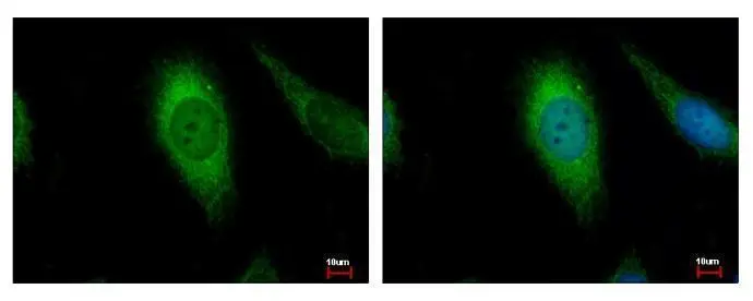
AIF antibody [N1N2], N-term detects AIFM1 protein at Mitochondria by immunofluorescent analysis. Sample: HeLa cells were fixed in 2% paraformaldehyde/culture medium at 37oC for 30 min. Green: AIFM1 protein stained by AIF antibody [N1N2], N-term (GTX102399) diluted at 1:500. Blue: Hoechst 33343 staining.
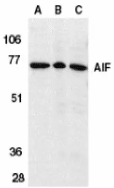
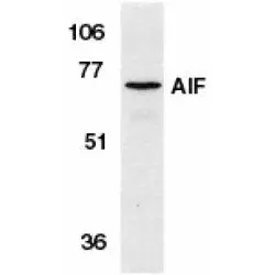
![AIF antibody [N1N2], N-term detects AIFM1 protein by Western blot analysis. A. 30 μg 293T whole cell lysate/extract B. 30 μg A431 whole cell lysate/extract C. 30 μg HeLa whole cell lysate/extract D. 30 μg HepG2 whole cell lysate/extract E. 30 μg A375 whole cell lysate/extract 7.5 % SDS-PAGE AIF antibody [N1N2], N-term (GTX102399) dilution: 1:1000](https://www.genetex.com/upload/website/prouct_img/normal/GTX102399/GTX102399_39925_WB_w_23060100_227.webp "AIF antibody [N1N2], N-term detects AIFM1 protein by Western blot analysis. A. 30 μg 293T whole cell lysate/extract B. 30 μg A431 whole cell lysate/extract C. 30 μg HeLa whole cell lysate/extract D. 30 μg HepG2 whole cell lysate/extract E. 30 μg A375 whole cell lysate/extract 7.5 % SDS-PAGE AIF antibody [N1N2], N-term (GTX102399) dilution: 1:1000")
 antibody at 1:100 dilution.
Antigen Retrieval: Trilogy? (EDTA based, pH 8.0) buffer, 15min")
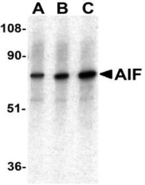
![Non-transfected (–) and transfected (+) 293T whole cell extracts (30 μg) were separated by 7.5% SDS-PAGE, and the membrane was blotted with AIF antibody [N1N2], N-term (GTX102399) diluted at 1:4000.](https://www.genetex.com/upload/website/prouct_img/normal/GTX102399/GTX102399_39925_20160804_WB_shRNA_watermark_w_23060100_700.webp "Non-transfected (–) and transfected (+) 293T whole cell extracts (30 μg) were separated by 7.5% SDS-PAGE, and the membrane was blotted with AIF antibody [N1N2], N-term (GTX102399) diluted at 1:4000.")
 antibody at 1:100 dilution.
Antigen Retrieval: Trilogy? (EDTA based, pH 8.0) buffer, 15min")
AIF antibody [N1N2], N-term detects AIFM1 protein at Mitochondria by immunofluorescent analysis. Sample: HeLa cells were fixed in 2% paraformaldehyde/culture medium at 37oC for 30 min. Green: AIFM1 protein stained by AIF antibody [N1N2], N-term (GTX102399) diluted at 1:500. Blue: Hoechst 33343 staining.
AIF antibody [N1N2], N-term
GTX102399
ApplicationsImmunoFluorescence, Western Blot, ImmunoCytoChemistry, ImmunoHistoChemistry, ImmunoHistoChemistry Paraffin
Product group Antibodies
ReactivityHuman
TargetAIFM1
Overview
- SupplierGeneTex
- Product NameAIF antibody [N1N2], N-term
- Delivery Days Customer9
- Application Supplier NoteWB: 1:500-1:10000. ICC/IF: 1:100-1:1000. IHC-P: 1:100-1:1000. *Optimal dilutions/concentrations should be determined by the researcher.Not tested in other applications.
- ApplicationsImmunoFluorescence, Western Blot, ImmunoCytoChemistry, ImmunoHistoChemistry, ImmunoHistoChemistry Paraffin
- CertificationResearch Use Only
- ClonalityPolyclonal
- Concentration0.72 mg/ml
- ConjugateUnconjugated
- Gene ID9131
- Target nameAIFM1
- Target descriptionapoptosis inducing factor mitochondria associated 1
- Target synonymsAIF, AUNX1, CMT2D, CMTX4, COWCK, COXPD6, DFNX5, NADMR, NAMSD, PDCD8, SEMDHL, apoptosis-inducing factor 1, mitochondrial, apoptosis-inducing factor, mitochondrion-associated, 1, auditory neuropathy, X-linked recessive 1, programmed cell death 8 (apoptosis-inducing factor), striatal apoptosis-inducing factor, testicular secretory protein Li 4
- HostRabbit
- IsotypeIgG
- Protein IDO95831
- Protein NameApoptosis-inducing factor 1, mitochondrial
- Scientific DescriptionThis gene encodes a flavoprotein essential for nuclear disassembly in apoptotic cells that is found in the mitochondrial intermembrane space in healthy cells. Induction of apoptosis results in the translocation of this protein to the nucleus where it effects chromosome condensation and fragmentation. In addition, this gene product induces mitochondria to release the apoptogenic proteins cytochrome c and caspase-9. Several alternative transcripts encoding different isoforms have been identified for this gene. [provided by RefSeq]
- ReactivityHuman
- Storage Instruction-20°C or -80°C,2°C to 8°C
- UNSPSC41116161
Datasheet
Related products

Product group Antibodies
Anti-AIFM1 AntibodyA97728
ApplicationsWestern Blot, ELISA
ReactivityHuman, Mouse, Rat
- SizePrice
Product group Antibodies
Anti-AIFM1 Antibody144-02568
ApplicationsImmunoFluorescence, ImmunoPrecipitation, Western Blot, ImmunoHistoChemistry
ReactivityHuman, Mouse
TargetAIFM1
- SizePrice
Product group Antibodies
References
AIF Polyclonal AntibodyBS-0037R
ApplicationsImmunoFluorescence, Western Blot, ELISA, ImmunoCytoChemistry, ImmunoHistoChemistry, ImmunoHistoChemistry Frozen, ImmunoHistoChemistry Paraffin
ReactivityBovine, Canine, Chicken, Human, Mouse, Porcine, Rabbit, Rat, Sheep
TargetAIFM1
- SizePrice
Product group Antibodies
AIFM1 AntibodyCSB-PA000836
ApplicationsImmunoFluorescence, Western Blot, ELISA, ImmunoHistoChemistry
ReactivityHuman, Mouse, Rat
TargetAIFM1
- SizePrice
Product group Antibodies

Goat anti-AIFM1 (aa183-195)EB12621
ApplicationsImmunoFluorescence, Western Blot, ELISA, ImmunoHistoChemistry
ReactivityBovine, Human, Mouse
TargetAIFM1
- SizePrice
Product group Antibodies
Aifm1 Polyclonal AntibodyCAC11738
ApplicationsImmunoFluorescence, Western Blot, ELISA, ImmunoHistoChemistry
TargetAIFM1
- SizePrice
Product group Antibodies
AIF antibodyGTX21998
ApplicationsWestern Blot, ELISA, ImmunoHistoChemistry, ImmunoHistoChemistry Paraffin
ReactivityHuman, Mouse, Rat
TargetAIFM1
- SizePrice
Product group Antibodies
AIF antibodyGTX21999
ApplicationsImmunoFluorescence, Western Blot, ImmunoCytoChemistry
ReactivityHuman
TargetAIFM1
- SizePrice
Product group Antibodies
AIF antibodyGTX22086
ApplicationsImmunoFluorescence, Western Blot, ImmunoCytoChemistry
ReactivityHuman, Mouse, Rat
TargetAIFM1
- SizePrice
![Various whole cell extracts (30 μg) were separated by 7.5% SDS-PAGE, and the membrane was blotted with AIF antibody [GT1143] (GTX00833) diluted at 1:1000. The HRP-conjugated anti-rabbit IgG antibody (GTX213110-01) was used to detect the primary antibody.](https://www.genetex.com/upload/website/prouct_img/normal/GTX00833/GTX00833_4000000015_20200410_WB_w_23053121_106.webp)
Product group Antibodies
AIF antibody [GT1143]GTX00833
ApplicationsWestern Blot, ImmunoHistoChemistry, ImmunoHistoChemistry Paraffin
ReactivityHuman, Mouse, Rat
TargetAIFM1
- SizePrice