

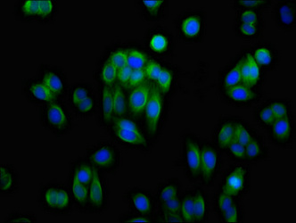
Immunofluorescence staining of HepG2 cells with CSB-PA001542LA01HU at 1:100, counter-stained with DAPI. The cells were fixed in 4% formaldehyde, permeabilized using 0.2% Triton X-100 and blocked in 10% normal Goat Serum. The cells were then incubated with the antibody overnight at 4°C. The secondary antibody was Alexa Fluor 488-congugated AffiniPure Goat Anti-Rabbit IgG(H+L).
Immunofluorescence staining of HepG2 cells with CSB-PA001542LA01HU at 1:100, counter-stained with DAPI. The cells were fixed in 4% formaldehyde, permeabilized using 0.2% Triton X-100 and blocked in 10% normal Goat Serum. The cells were then incubated with the antibody overnight at 4°C. The secondary antibody was Alexa Fluor 488-congugated AffiniPure Goat Anti-Rabbit IgG(H+L).
AKR1C1 Antibody
CSB-PA001542LA01HU
ApplicationsImmunoFluorescence, ELISA
Product group Antibodies
ReactivityHuman
TargetAKR1C1
Overview
- SupplierCusabio
- Product NameAKR1C1 Antibody
- Delivery Days Customer20
- ApplicationsImmunoFluorescence, ELISA
- CertificationResearch Use Only
- ClonalityPolyclonal
- ConjugateUnconjugated
- Gene ID1645
- Target nameAKR1C1
- Target descriptionaldo-keto reductase family 1 member C1
- Target synonyms2-ALPHA-HSD, 20-ALPHA-HSD, C9, DD1, DD1/DD2, DDH, DDH1, H-37, HAKRC, HBAB, MBAB, aldo-keto reductase family 1 member C1, 20 alpha-hydroxysteroid dehydrogenase, aldo-keto reductase C, chlordecone reductase homolog HAKRC, dihydrodiol dehydrogenase 1, dihydrodiol dehydrogenase 1/2, dihydrodiol dehydrogenase 1; 20-alpha (3-alpha)-hydroxysteroid dehydrogenase, hepatic dihydrodiol dehydrogenase, high-affinity hepatic bile acid-binding protein, indanol dehydrogenase, trans-1,2-dihydrobenzene-1,2-diol dehydrogenase, type II 3-alpha-hydroxysteroid dehydrogenase
- HostRabbit
- IsotypeIgG
- Protein IDQ04828
- Protein NameAldo-keto reductase family 1 member C1
- Scientific DescriptionConverts progesterone to its inactive form, 20-alpha-dihydroxyprogesterone (20-alpha-OHP). In the liver and intestine, may have a role in the transport of bile. May have a role in monitoring the intrahepatic bile acid concentration. Has a low bile-binding ability. May play a role in myelin formation.
- ReactivityHuman
- Storage Instruction-20°C or -80°C
- UNSPSC41116161
Related products
Product group Antibodies
Anti-AKR1C1 AntibodyA28765
ApplicationsWestern Blot
ReactivityHuman, Mouse, Rat
- SizePrice
Product group Antibodies
Anti-AKR1C1 Antibody144-60143
ApplicationsWestern Blot
ReactivityHuman, Mouse
TargetAKR1C1
- SizePrice
Product group Antibodies
AKR1C1/2 Recombinant AntibodyBSM-62246R
ApplicationsFlow Cytometry, Western Blot
ReactivityHuman, Mouse, Rat
TargetAKR1C1
- SizePrice
Product group Antibodies

DDH / AKR1C1 AntibodyLS-C402612
ApplicationsWestern Blot, ELISA, ImmunoHistoChemistry
ReactivityHuman
TargetAKR1C1
- SizePrice

Product group Antibodies
Anti-AKR1C1 AntibodyHPA068265
ApplicationsWestern Blot, ImmunoCytoChemistry
ReactivityHuman
TargetAKR1C1
- SizePrice
Product group Antibodies
AKR1C1 antibodyGTX105620
ApplicationsImmunoFluorescence, Western Blot, ImmunoCytoChemistry, ImmunoHistoChemistry, ImmunoHistoChemistry Paraffin
ReactivityHuman, Mouse
TargetAKR1C1
- SizePrice
Product group Antibodies
Anti-AKR1C1Y058805
ApplicationsWestern Blot, ImmunoHistoChemistry
ReactivityHuman
- SizePrice

Product group Antibodies
Anti-AKR1C1/C2 Antibody Picoband(r)PB10036-CARRIER-FREE
ApplicationsFlow Cytometry, ImmunoFluorescence, Western Blot, ImmunoCytoChemistry
ReactivityHuman, Mouse, Rat
TargetAKR1C1
- SizePrice