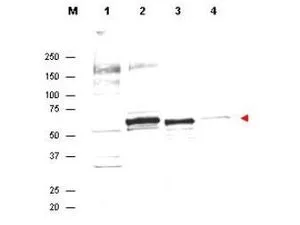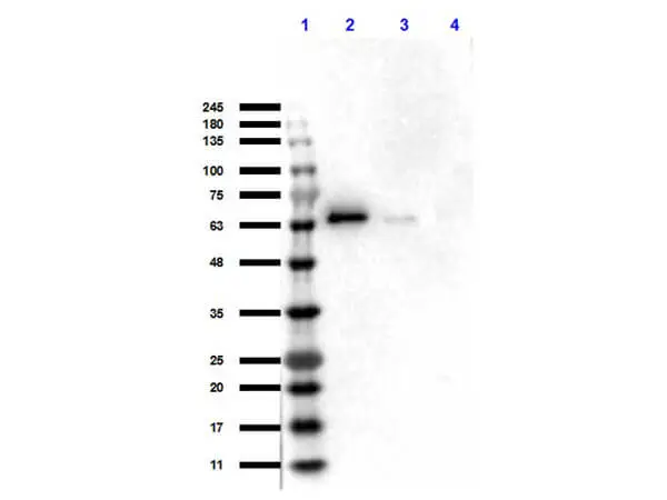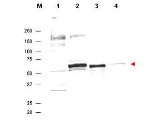
Anti-AKT Antibody in western blot showing detection of endogenous protein in whole cell extracts from H2O2 treated MCF7 cells (lane 2). Recombinant AKT is detected in lane 3. Lanes 1 and 4 contain wcl from HeLa and A431 cells, respectively. Specific band staining is completely blocked when antibody is pre-incubated with the immunizing peptide (data not shown). The band at ~55-60 kDa, indicated by the arrowhead, corresponds to the expected molecular weight of AKT. AKT antibody was diluted 1:300 in 1% BLOTTO and reacted with the membrane overnight at 4o C. IRDYE800 conjugated Dnky-anti-Sheep IgG was used at a 1:10,000 dilution for 45 min at room temperature.
AKT antibody
GTX48651
ApplicationsFlow Cytometry, ImmunoFluorescence, Western Blot, ELISA, ImmunoCytoChemistry, ImmunoHistoChemistry
Product group Antibodies
ReactivityHuman
Overview
- SupplierGeneTex
- Product NameAKT antibody
- Delivery Days Customer9
- Application Supplier NoteWB: 1:500-1:2000. ELISA: 1:5000-1:20000. *Optimal dilutions/concentrations should be determined by the researcher.Not tested in other applications.
- ApplicationsFlow Cytometry, ImmunoFluorescence, Western Blot, ELISA, ImmunoCytoChemistry, ImmunoHistoChemistry
- CertificationResearch Use Only
- ClonalityPolyclonal
- Concentration0.9 mg/ml
- ConjugateUnconjugated
- HostSheep
- IsotypeIgG
- Scientific DescriptionAKT is a component of the PI-3 kinase pathway and is activated by phosphorylation at Ser 473 and Thr 308. AKT is a cytoplasmic protein also known as Protein Kinase B (PKB) and rac (related to A and C kinases). AKT is a key regulator of many signal transduction pathways. AKT Exhibits tight control over cell proliferation and cell viability.Overexpression or inappropriate activation of AKT is noted in many types of cancer. AKT mediates many of the downstream events of PI 3-kinase (a lipid kinase activated by growth factors, cytokines and insulin). PI 3-kinase recruits AKT to the membrane, where it is activated by PDK1 phosphorylation. Once phosphorylated, AKT dissociates from the membrane and phosphorylates targets in the cytoplasm and the cell nucleus. AKT has two main roles: (i) inhibition of apoptosis; (ii) promotion of proliferation.
- ReactivityHuman
- Storage Instruction-20°C or -80°C,2°C to 8°C
- UNSPSC41116161


