

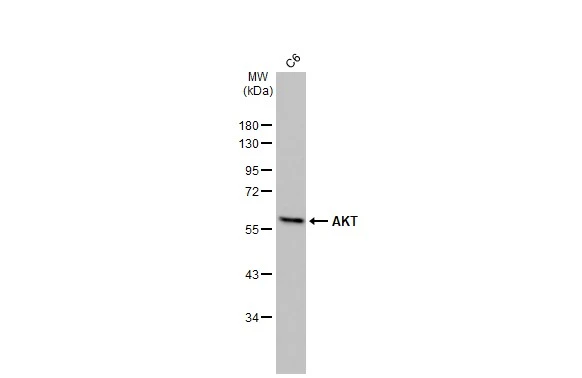
Whole cell extract (30 μg) was separated by 10% SDS-PAGE, and the membrane was blotted with AKT antibody [HL2915] (GTX640258) diluted at 1:1000. The HRP-conjugated anti-rabbit IgG antibody (GTX213110-01) was used to detect the primary antibody.
![AKT antibody [HL2915] detects AKT protein by immunofluorescent analysis. Sample: HeLa cells were fixed in ice-cold MeOH for 5 min. Green: AKT stained by AKT antibody [HL2915] (GTX640258) diluted at 1:500. Red: alpha Tubulin, a cytoskeleton marker, stained by alpha Tubulin antibody [GT114] (GTX628802) diluted at 1:1000. Blue: Fluoroshield with DAPI (GTX30920).](https://www.genetex.com/upload/website/prouct_img/normal/GTX640258/GTX640258_T-45390_20240510_ICC_IF_24052202_426.webp "AKT antibody [HL2915] detects AKT protein by immunofluorescent analysis. Sample: HeLa cells were fixed in ice-cold MeOH for 5 min. Green: AKT stained by AKT antibody [HL2915] (GTX640258) diluted at 1:500. Red: alpha Tubulin, a cytoskeleton marker, stained by alpha Tubulin antibody [GT114] (GTX628802) diluted at 1:1000. Blue: Fluoroshield with DAPI (GTX30920).")
![Whole cell extract (30 μg) was separated by 10% SDS-PAGE, and the membrane was blotted with AKT antibody [HL2915] (GTX640258) diluted at 1:1000. The HRP-conjugated anti-rabbit IgG antibody (GTX213110-01) was used to detect the primary antibody.](https://www.genetex.com/upload/website/prouct_img/normal/GTX640258/GTX640258_T-45390_20240531_WB_24060619_187.webp "Whole cell extract (30 μg) was separated by 10% SDS-PAGE, and the membrane was blotted with AKT antibody [HL2915] (GTX640258) diluted at 1:1000. The HRP-conjugated anti-rabbit IgG antibody (GTX213110-01) was used to detect the primary antibody.")
![AKT antibody [HL2915] detects AKT protein by immunohistochemical analysis. Sample: Paraffin-embedded rat esophagus. AKT stained by AKT antibody [HL2915] (GTX640258) diluted at 1:100. Antigen Retrieval: Citrate buffer, pH 6.0, 15 min](https://www.genetex.com/upload/website/prouct_img/normal/GTX640258/GTX640258_T-45390_20240619_IHC-P_R_24062501_570.webp "AKT antibody [HL2915] detects AKT protein by immunohistochemical analysis. Sample: Paraffin-embedded rat esophagus. AKT stained by AKT antibody [HL2915] (GTX640258) diluted at 1:100. Antigen Retrieval: Citrate buffer, pH 6.0, 15 min")
![AKT antibody [HL2915] detects AKT protein by immunohistochemical analysis. Sample: Paraffin-embedded human breast carcinoma. AKT stained by AKT antibody [HL2915] (GTX640258) diluted at 1:100. Antigen Retrieval: Citrate buffer, pH 6.0, 15 min](https://www.genetex.com/upload/website/prouct_img/normal/GTX640258/GTX640258_T-45390_20240619_IHC-P_24062501_700.webp "AKT antibody [HL2915] detects AKT protein by immunohistochemical analysis. Sample: Paraffin-embedded human breast carcinoma. AKT stained by AKT antibody [HL2915] (GTX640258) diluted at 1:100. Antigen Retrieval: Citrate buffer, pH 6.0, 15 min")
![AKT antibody [HL2915] detects AKT protein by immunohistochemical analysis. Sample: Paraffin-embedded mouse tissues. AKT stained by AKT antibody [HL2915] (GTX640258) diluted at 1:100. Antigen Retrieval: Citrate buffer, pH 6.0, 15 min](https://www.genetex.com/upload/website/prouct_img/normal/GTX640258/GTX640258_T-45390_20240619_IHC-P_multiple_M_24062501_370.webp "AKT antibody [HL2915] detects AKT protein by immunohistochemical analysis. Sample: Paraffin-embedded mouse tissues. AKT stained by AKT antibody [HL2915] (GTX640258) diluted at 1:100. Antigen Retrieval: Citrate buffer, pH 6.0, 15 min")
![Non-transfected (–) and transfected (+) 293T whole cell extracts were separated by 10% SDS-PAGE, and the membrane was blotted with AKT antibody [HL2915] (GTX640258) diluted at 1:5000. The HRP-conjugated anti-rabbit IgG antibody (GTX213110-01) was used to detect the primary antibody.](https://www.genetex.com/upload/website/prouct_img/normal/GTX640258/GTX640258_45446_20240920_WB_multiple_B_24092600_500.webp "Non-transfected (–) and transfected (+) 293T whole cell extracts were separated by 10% SDS-PAGE, and the membrane was blotted with AKT antibody [HL2915] (GTX640258) diluted at 1:5000. The HRP-conjugated anti-rabbit IgG antibody (GTX213110-01) was used to detect the primary antibody.")
![Various whole cell extracts (30 μg) were separated by 10% SDS-PAGE, and the membrane was blotted with AKT antibody [HL2915] (GTX640258) diluted at 1:1000. The HRP-conjugated anti-rabbit IgG antibody (GTX213110-01) was used to detect the primary antibody.](https://www.genetex.com/upload/website/prouct_img/normal/GTX640258/GTX640258_45446_20250411_WB_M_25041720_346.webp "Various whole cell extracts (30 μg) were separated by 10% SDS-PAGE, and the membrane was blotted with AKT antibody [HL2915] (GTX640258) diluted at 1:1000. The HRP-conjugated anti-rabbit IgG antibody (GTX213110-01) was used to detect the primary antibody.")
Whole cell extract (30 μg) was separated by 10% SDS-PAGE, and the membrane was blotted with AKT antibody [HL2915] (GTX640258) diluted at 1:1000. The HRP-conjugated anti-rabbit IgG antibody (GTX213110-01) was used to detect the primary antibody.
AKT antibody [HL2915]
GTX640258
ApplicationsImmunoFluorescence, Western Blot, ImmunoCytoChemistry, ImmunoHistoChemistry, ImmunoHistoChemistry Paraffin
Product group Antibodies
ReactivityHuman, Mouse, Rat
TargetAKT1
Overview
- SupplierGeneTex
- Product NameAKT antibody [HL2915]
- Delivery Days Customer7
- Application Supplier NoteWB: 1:500-1:10000. *Optimal dilutions/concentrations should be determined by the researcher.Not tested in other applications.
- ApplicationsImmunoFluorescence, Western Blot, ImmunoCytoChemistry, ImmunoHistoChemistry, ImmunoHistoChemistry Paraffin
- CertificationResearch Use Only
- ClonalityMonoclonal
- Clone IDHL2915
- Concentration1 mg/ml
- ConjugateUnconjugated
- Gene ID207
- Target nameAKT1
- Target descriptionAKT serine/threonine kinase 1
- Target synonymsAKT, PKB, PKB-ALPHA, PRKBA, RAC, RAC-ALPHA, RAC-alpha serine/threonine-protein kinase, AKT1m, PKB alpha, RAC-PK-alpha, protein kinase B alpha, proto-oncogene c-Akt, rac protein kinase alpha, serine-threonine protein kinase, v-akt murine thymoma viral oncogene homolog 1, v-akt murine thymoma viral oncogene-like protein 1
- HostRabbit
- IsotypeIgG
- Protein IDP31749
- Protein NameRAC-alpha serine/threonine-protein kinase
- ReactivityHuman, Mouse, Rat
- Storage Instruction-20°C or -80°C,2°C to 8°C
- UNSPSC41116161
Datasheet
Related products
Product group Antibodies
Anti-AKT1 AntibodyA285951
ApplicationsWestern Blot, ELISA
ReactivityHuman, Mouse
- SizePrice
Product group Antibodies
Anti-AKT1 Antibody144-11016
ApplicationsImmunoFluorescence, Western Blot, ImmunoHistoChemistry
ReactivityHuman, Mouse, Rat
TargetAKT1
- SizePrice
Product group Antibodies

Anti-pAKT [AbAb-pAKT]AB04102-10.0
ApplicationsImmunoPrecipitation, Western Blot, ELISA
ReactivityHuman, Mouse
TargetAKT1
- SizePrice

Product group Antibodies
Anti-AKT1 AntibodyAMAB90834
ApplicationsWestern Blot, ImmunoCytoChemistry
ReactivityHuman
TargetAKT1
- SizePrice
Product group Antibodies

AKT1 AntibodyLS-C831478
ApplicationsImmunoHistoChemistry
ReactivityHuman
TargetAKT1
- SizePrice

Product group Antibodies
Anti-AKT1,2,3 Antibody Picoband(r)A00024-2-CARRIER-FREE
ApplicationsFlow Cytometry, ImmunoFluorescence, Western Blot, ELISA, ImmunoCytoChemistry
ReactivityHuman, Mouse, Rat
TargetAKT1
- SizePrice
Product group Antibodies
AKT1 Polyclonal AntibodyBS-0115M
ApplicationsImmunoFluorescence, Western Blot, ELISA, ImmunoCytoChemistry, ImmunoHistoChemistry, ImmunoHistoChemistry Frozen, ImmunoHistoChemistry Paraffin
ReactivityBovine, Canine, Chicken, Human, Mouse, Porcine, Rabbit, Rat, Sheep
TargetAKT1
- SizePrice
Product group Antibodies
AKT1/AKT2/AKT3 AntibodyCSB-PA000848
ApplicationsWestern Blot, ELISA, ImmunoHistoChemistry
ReactivityHuman, Mouse, Rat
TargetAKT1
- SizePrice
Product group Antibodies

Goat anti-AKT1EB06875
ApplicationsWestern Blot, ELISA
ReactivityBovine, Human, Mouse
TargetAKT1
- SizePrice