


Cross-reactivity assessment of AKT1 Mouse Monoclonal Antibody AE00130 (1ug/ml) on CDI’s Protein Array containing more than 19,000 full-length human proteins.
 and intact heavy and light chains at 50kDa and 28kDa resp. ®")
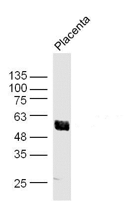
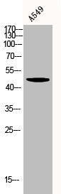
 stained with AKT1 Mouse Monoclonal Antibody AE00130 at 1-2ug/ml (1h at ambient temp). ECL staining by HRP.")

Cross-reactivity assessment of AKT1 Mouse Monoclonal Antibody AE00130 (1ug/ml) on CDI’s Protein Array containing more than 19,000 full-length human proteins.
AKT1 Monoclonal Antibody AE00130
AE00130
TargetAKT1
Product group Antibodies
Product AE00130 is not available
Product not available
There may be an alternative product available, please contact our technical support team.
Overview
- SupplierAeonian Biotech
- Product NameAKT1 Monoclonal Antibody AE00130
- Delivery Days Customer9
- Applications SupplierIHC, PA, WB
- CertificationResearch Use Only
- ClonalityMonoclonal
- Clone IDAKT1/2552
- ConjugateUnconjugated
- Gene ID207
- Target nameAKT1
- Target descriptionAKT serine/threonine kinase 1
- Target synonymsAKT, PKB, PKB-ALPHA, PRKBA, RAC, RAC-ALPHA, RAC-alpha serine/threonine-protein kinase, AKT1m, PKB alpha, RAC-PK-alpha, protein kinase B alpha, proto-oncogene c-Akt, rac protein kinase alpha, serine-threonine protein kinase, v-akt murine thymoma viral oncogene homolog 1, v-akt murine thymoma viral oncogene-like protein 1
- HostMouse
- IsotypeIgG2b kappa
- Protein IDP31749
- Protein NameRAC-alpha serine/threonine-protein kinase
- Scientific DescriptionAKT1 Monoclonal Antibody AE00130
- Shelf life instructionIntegrity warranted for 24 months after purchase when handled and stored according to instructions, see below.
- Reactivity SupplierHuman
- Storage Instruction2-8°C
- UNSPSC12352203
Datasheet
MSDS
Related products
Product group Antibodies
Anti-AKT1 AntibodyA285951
ApplicationsWestern Blot, ELISA
ReactivityHuman, Mouse
- SizePrice
Product group Antibodies
Anti-AKT1 Antibody144-11016
ApplicationsImmunoFluorescence, Western Blot, ImmunoHistoChemistry
ReactivityHuman, Mouse, Rat
TargetAKT1
- SizePrice
Product group Antibodies

Anti-pAKT [AbAb-pAKT]AB04102-10.0
ApplicationsImmunoPrecipitation, Western Blot, ELISA
ReactivityHuman, Mouse
TargetAKT1
- SizePrice

Product group Antibodies
Anti-AKT1 AntibodyAMAB90834
ApplicationsWestern Blot, ImmunoCytoChemistry
ReactivityHuman
TargetAKT1
- SizePrice
Product group Antibodies

AKT1 AntibodyLS-C831478
ApplicationsImmunoHistoChemistry
ReactivityHuman
TargetAKT1
- SizePrice

Product group Antibodies
Anti-AKT1,2,3 Antibody Picoband(r)A00024-2-CARRIER-FREE
ApplicationsFlow Cytometry, ImmunoFluorescence, Western Blot, ELISA, ImmunoCytoChemistry
ReactivityHuman, Mouse, Rat
TargetAKT1
- SizePrice
Product group Antibodies
AKT1 Polyclonal AntibodyBS-0115M
ApplicationsImmunoFluorescence, Western Blot, ELISA, ImmunoCytoChemistry, ImmunoHistoChemistry, ImmunoHistoChemistry Frozen, ImmunoHistoChemistry Paraffin
ReactivityBovine, Canine, Chicken, Human, Mouse, Porcine, Rabbit, Rat, Sheep
TargetAKT1
- SizePrice
Product group Antibodies
AKT1/AKT2/AKT3 AntibodyCSB-PA000848
ApplicationsWestern Blot, ELISA, ImmunoHistoChemistry
ReactivityHuman, Mouse, Rat
TargetAKT1
- SizePrice
Product group Antibodies

Goat anti-AKT1EB06875
ApplicationsWestern Blot, ELISA
ReactivityBovine, Human, Mouse
TargetAKT1
- SizePrice