

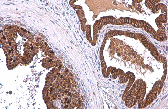
ALDH1A3 antibody [N2C2], Internal detects ALDH1A3 protein at cytoplasm by immunohistochemical analysis. Sample: Paraffin-embedded mouse prostate. ALDH1A3 stained by ALDH1A3 antibody [N2C2], Internal (GTX110784) diluted at 1:500. Antigen Retrieval: Citrate buffer, pH 6.0, 15 min
![ALDH1A3 antibody [N2C2], Internal detects ALDH1A3 protein at cytoplasm by immunohistochemical analysis. Sample: Paraffin-embedded rat prostate. ALDH1A3 stained by ALDH1A3 antibody [N2C2], Internal (GTX110784) diluted at 1:500. Antigen Retrieval: Citrate buffer, pH 6.0, 15 min](https://www.genetex.com/upload/website/prouct_img/normal/GTX110784/GTX110784_43054_20181221_IHC-P_R_w_23060500_141.webp "ALDH1A3 antibody [N2C2], Internal detects ALDH1A3 protein at cytoplasm by immunohistochemical analysis. Sample: Paraffin-embedded rat prostate. ALDH1A3 stained by ALDH1A3 antibody [N2C2], Internal (GTX110784) diluted at 1:500. Antigen Retrieval: Citrate buffer, pH 6.0, 15 min")
![Various whole cell extracts (30 μg) were separated by 10% SDS-PAGE, and the membrane was blotted with ALDH1A3 antibody [N2C2], Internal (GTX110784) diluted at 1:500. The HRP-conjugated anti-rabbit IgG antibody (GTX213110-01) was used to detect the primary antibody. Corresponding RNA expression data for the same cell lines are based on Human Protein Atlas program.](https://www.genetex.com/upload/website/prouct_img/normal/GTX110784/GTX110784_43082_20191025_WB_TPM_watermark_w_23060500_574.webp "Various whole cell extracts (30 μg) were separated by 10% SDS-PAGE, and the membrane was blotted with ALDH1A3 antibody [N2C2], Internal (GTX110784) diluted at 1:500. The HRP-conjugated anti-rabbit IgG antibody (GTX213110-01) was used to detect the primary antibody. Corresponding RNA expression data for the same cell lines are based on Human Protein Atlas program.")
![ALDH1A3 antibody [N2C2], Internal detects ALDH1A3 protein at cytoplasm by immunofluorescent analysis. Sample: A431 cells were fixed in 4% paraformaldehyde at RT for 15 min. Green: ALDH1A3 stained by ALDH1A3 antibody [N2C2], Internal (GTX110784) diluted at 1:500. Blue: Hoechst 33342 staining. Scale bar= 10μm.](https://www.genetex.com/upload/website/prouct_img/normal/GTX110784/GTX110784_43082_20180314_ICC_IF_w_23060500_285.webp "ALDH1A3 antibody [N2C2], Internal detects ALDH1A3 protein at cytoplasm by immunofluorescent analysis. Sample: A431 cells were fixed in 4% paraformaldehyde at RT for 15 min. Green: ALDH1A3 stained by ALDH1A3 antibody [N2C2], Internal (GTX110784) diluted at 1:500. Blue: Hoechst 33342 staining. Scale bar= 10μm.")
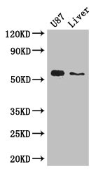
![Whole cell extract (30 μg) was separated by 10% SDS-PAGE, and the membrane was blotted with ALDH1A3 antibody [N2C2], Internal (GTX110784) diluted at 1:1000. The HRP-conjugated anti-rabbit IgG antibody (GTX213110-01) was used to detect the primary antibody.](https://www.genetex.com/upload/website/prouct_img/normal/GTX110784/GTX110784_42992_20171006_WB_w_23060500_115.webp "Whole cell extract (30 μg) was separated by 10% SDS-PAGE, and the membrane was blotted with ALDH1A3 antibody [N2C2], Internal (GTX110784) diluted at 1:1000. The HRP-conjugated anti-rabbit IgG antibody (GTX213110-01) was used to detect the primary antibody.")
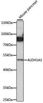
![Whole cell extract (30 μg) was separated by 7.5% SDS-PAGE, and the membrane was blotted with ALDH1A3 antibody [N2C2], Internal (GTX110784) diluted at 1:2000.](https://www.genetex.com/upload/website/prouct_img/normal/GTX110784/GTX110784_41605_20170209_WB_w_23060500_832.webp "Whole cell extract (30 μg) was separated by 7.5% SDS-PAGE, and the membrane was blotted with ALDH1A3 antibody [N2C2], Internal (GTX110784) diluted at 1:2000.")
ALDH1A3 antibody [N2C2], Internal detects ALDH1A3 protein at cytoplasm by immunohistochemical analysis. Sample: Paraffin-embedded mouse prostate. ALDH1A3 stained by ALDH1A3 antibody [N2C2], Internal (GTX110784) diluted at 1:500. Antigen Retrieval: Citrate buffer, pH 6.0, 15 min
ALDH1A3 antibody [N2C2], Internal
GTX110784
ApplicationsImmunoFluorescence, Western Blot, ImmunoCytoChemistry, ImmunoHistoChemistry, ImmunoHistoChemistry Paraffin
Product group Antibodies
ReactivityHuman, Mouse, Rat
TargetALDH1A3
Overview
- SupplierGeneTex
- Product NameALDH1A3 antibody [N2C2], Internal
- Delivery Days Customer9
- Application Supplier NoteWB: 1:500-1:3000. ICC/IF: 1:100-1:1000. IHC-P: 1:100-1:1000. *Optimal dilutions/concentrations should be determined by the researcher.Not tested in other applications.
- ApplicationsImmunoFluorescence, Western Blot, ImmunoCytoChemistry, ImmunoHistoChemistry, ImmunoHistoChemistry Paraffin
- CertificationResearch Use Only
- ClonalityPolyclonal
- Concentration0.1 mg/ml
- ConjugateUnconjugated
- Gene ID220
- Target nameALDH1A3
- Target descriptionaldehyde dehydrogenase 1 family member A3
- Target synonymsALDH1A6, ALDH6, MCOP8, RALDH3, retinaldehyde dehydrogenase 3, acetaldehyde dehydrogenase 6, aldehyde dehydrogenase 6
- HostRabbit
- IsotypeIgG
- Protein IDP47895
- Protein NameRetinaldehyde dehydrogenase 3
- Scientific DescriptionAldehyde dehydrogenase isozymes are thought to play a major role in the detoxification of aldehydes generated by alcohol metabolism and lipid peroxidation. The enzyme encoded by this gene uses retinal as a substrate, either in a free or cellular retinol-binding protein form. [provided by RefSeq]
- ReactivityHuman, Mouse, Rat
- Storage Instruction-20°C or -80°C,2°C to 8°C
- UNSPSC41116161
Datasheet
Related products

Product group Antibodies
Anti-ALDH1A3 Antibody Picoband(r)A03030-CARRIER-FREE
ApplicationsFlow Cytometry, ImmunoFluorescence, Western Blot, ImmunoCytoChemistry, ImmunoHistoChemistry, ImmunoHistoChemistry Frozen
ReactivityHuman, Mouse, Rat
TargetALDH1A3
- SizePrice
Product group Antibodies
Anti-ALDH1A3 Antibody144-63650
ApplicationsWestern Blot
ReactivityHuman, Mouse
TargetALDH1A3
- SizePrice

Product group Antibodies
Anti-ALDH1A3 AntibodyAMAB91754
ApplicationsWestern Blot, ImmunoCytoChemistry, ImmunoHistoChemistry
ReactivityHuman
TargetALDH1A3
- SizePrice
Product group Antibodies
Anti-ALDH1A3 AntibodyA90205
ApplicationsWestern Blot
ReactivityHuman, Mouse
- SizePrice
Product group Antibodies

ALDH1A3 AntibodyLS-C750201
ApplicationsWestern Blot
ReactivityHuman, Mouse
TargetALDH1A3
- SizePrice
Product group Antibodies
ALDH1A3 Polyclonal AntibodyBS-25689R
ApplicationsImmunoHistoChemistry
ReactivityBovine, Canine, Chicken, Equine, Human, Mouse, Porcine, Rabbit, Rat, Sheep
TargetALDH1A3
- SizePrice
Product group Antibodies
ALDH1A3 AntibodyCSB-PA001567LA01HU
ApplicationsImmunoFluorescence, Western Blot, ELISA
ReactivityHuman, Mouse
TargetALDH1A3
- SizePrice
Product group Antibodies
Aldh1A3 Polyclonal AntibodyCAC08120
ApplicationsImmunoFluorescence, Western Blot, ELISA
ReactivityMouse
TargetALDH1A3
- SizePrice
![Whole cell extract (30 μg) was separated by 10% SDS-PAGE, and the membrane was blotted with ALDH1A3 antibody [GT926] (GTX633822) diluted at 1:1000. The HRP-conjugated anti-mouse IgG antibody (GTX213111-01) was used to detect the primary antibody.](https://www.genetex.com/upload/website/prouct_img/normal/GTX633822/GTX633822_42688_20171006_WB_2_w_23061202_702.webp)
Product group Antibodies
ALDH1A3 antibody [GT926]GTX633822
ApplicationsImmunoFluorescence, Western Blot, ImmunoCytoChemistry, ImmunoHistoChemistry, ImmunoHistoChemistry Paraffin
ReactivityHuman, Mouse, Rat
TargetALDH1A3
- SizePrice