

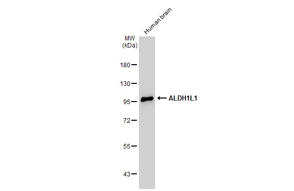
Human tissue extract (30 μg) was separated by 7.5% SDS-PAGE, and the membrane was blotted with ALDH1L1 antibody (GTX131047) diluted at 1:1000. The HRP-conjugated anti-rabbit IgG antibody (GTX213110-01) was used to detect the primary antibody.
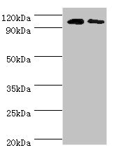
 was separated by 7.5% SDS-PAGE, and the membrane was blotted with ALDH1L1 antibody (GTX131047) diluted at 1:5000. The HRP-conjugated anti-rabbit IgG antibody (GTX213110-01) was used to detect the primary antibody.")
 were separated by 7.5% SDS-PAGE, and the membrane was blotted with ALDH1L1 antibody (GTX131047) diluted at 1:500. The HRP-conjugated anti-rabbit IgG antibody (GTX213110-01) was used to detect the primary antibody.")
 was separated by 7.5% SDS-PAGE, and the membrane was blotted with ALDH1L1 antibody (GTX131047) diluted at 1:5000. The HRP-conjugated anti-rabbit IgG antibody (GTX213110-01) was used to detect the primary antibody.")
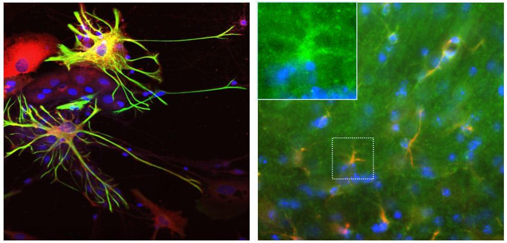
![ALDH1L1 antibody detects ALDH1L1 protein by immunohistochemical analysis. Sample: Paraffin-embedded mouse hippocampus. Green: ALDH1L1 stained by ALDH1L1 antibody (GTX131047) diluted at 1:500. Red: GFAP antibody [GT25] (GTX641104) diluted at 1:250. Blue: Fluoroshield with DAPI (GTX30920). Antigen Retrieval: Citrate buffer, pH 6.0, 15 min](https://www.genetex.com/upload/website/prouct_img/normal/GTX131047/GTX131047_45778_20251106_IHC-P_M_25111401_978.webp "ALDH1L1 antibody detects ALDH1L1 protein by immunohistochemical analysis. Sample: Paraffin-embedded mouse hippocampus. Green: ALDH1L1 stained by ALDH1L1 antibody (GTX131047) diluted at 1:500. Red: GFAP antibody [GT25] (GTX641104) diluted at 1:250. Blue: Fluoroshield with DAPI (GTX30920). Antigen Retrieval: Citrate buffer, pH 6.0, 15 min")
Human tissue extract (30 μg) was separated by 7.5% SDS-PAGE, and the membrane was blotted with ALDH1L1 antibody (GTX131047) diluted at 1:1000. The HRP-conjugated anti-rabbit IgG antibody (GTX213110-01) was used to detect the primary antibody.
ALDH1L1 antibody
GTX131047
ApplicationsWestern Blot, ImmunoHistoChemistry, ImmunoHistoChemistry Paraffin
Product group Antibodies
ReactivityHuman, Mouse, Rat
TargetALDH1L1
Overview
- SupplierGeneTex
- Product NameALDH1L1 antibody
- Delivery Days Customer9
- Application Supplier NoteWB: 1:500-1:10000. *Optimal dilutions/concentrations should be determined by the researcher.Not tested in other applications.
- ApplicationsWestern Blot, ImmunoHistoChemistry, ImmunoHistoChemistry Paraffin
- CertificationResearch Use Only
- ClonalityPolyclonal
- Concentration0.82 mg/ml
- ConjugateUnconjugated
- Gene ID10840
- Target nameALDH1L1
- Target descriptionaldehyde dehydrogenase 1 family member L1
- Target synonyms10-FTHFDH, 10-fTHF, FDH, FTHFD, cytosolic 10-formyltetrahydrofolate dehydrogenase, 10-formyltetrahydrofolate dehydrogenase, MER57B-ALDH1L1
- HostRabbit
- IsotypeIgG
- Protein IDO75891
- Protein NameCytosolic 10-formyltetrahydrofolate dehydrogenase
- Scientific DescriptionThe protein encoded by this gene catalyzes the conversion of 10-formyltetrahydrofolate, NADP, and water to tetrahydrofolate, NADPH, and carbon dioxide. The encoded protein belongs to the aldehyde dehydrogenase family and is responsible for formate oxidation in vivo. Deficiencies in this gene can result in an accumulation of formate and subsequent methanol poisoning. [provided by RefSeq]
- ReactivityHuman, Mouse, Rat
- Storage Instruction-20°C or -80°C,2°C to 8°C
- UNSPSC41116161
Datasheet
Related products
Product group Antibodies
ALDH1L1 AntibodyCSB-PA001569ESR1HU
ApplicationsWestern Blot, ELISA, ImmunoHistoChemistry
ReactivityHuman, Mouse
TargetALDH1L1
- SizePrice
Product group Antibodies
Anti-ALDH1L1 AntibodyA85310
ApplicationsImmunoFluorescence, Western Blot, ImmunoCytoChemistry
ReactivityHuman, Mouse, Rat
- SizePrice

Product group Antibodies
Anti-ALDH1L1 AntibodyHPA036900
ApplicationsWestern Blot, ImmunoHistoChemistry
ReactivityHuman
TargetALDH1L1
- SizePrice

Product group Antibodies
Anti-ALDH1L1 Antibody Picoband(r)A04615-1-CARRIER-FREE
ApplicationsFlow Cytometry, ImmunoFluorescence, Western Blot, ELISA, ImmunoCytoChemistry, ImmunoHistoChemistry
ReactivityHuman, Monkey, Mouse, Rat
TargetALDH1L1
- SizePrice
Product group Antibodies

ALDH1L1 AntibodyLS-C349022
ApplicationsWestern Blot, ImmunoHistoChemistry
ReactivityHuman, Mouse, Rat
TargetALDH1L1
- SizePrice
![IHC-P analysis of human bladder carcinoma tissue using GTX84891 ALDH1L1 antibody [6A10].](https://www.genetex.com/upload/website/prouct_img/normal/GTX84891/GTX84891_3219_IHC-P_w_23061420_829.webp)
Product group Antibodies
ALDH1L1 antibody [6A10]GTX84891
ApplicationsFlow Cytometry, Western Blot, ImmunoHistoChemistry, ImmunoHistoChemistry Paraffin
ReactivityCanine, Human
TargetALDH1L1
- SizePrice
![FACS analysis of HeLa cells using GTX84892 ALDH1L1 antibody [5G8]. Red : Primary antibody Blue : Negative control antibody](https://www.genetex.com/upload/website/prouct_img/normal/GTX84892/GTX84892_606_FACS_w_23061420_106.webp)
Product group Antibodies
ALDH1L1 antibody [5G8]GTX84892
ApplicationsFlow Cytometry, ImmunoFluorescence, Western Blot, ImmunoCytoChemistry, ImmunoHistoChemistry, ImmunoHistoChemistry Paraffin
ReactivityCanine, Human
TargetALDH1L1
- SizePrice
![ICC/IF analysis of COS7 cells transiently transfected with ALDH1L1 plasmid using GTX84894 ALDH1L1 antibody [7G6].](https://www.genetex.com/upload/website/prouct_img/normal/GTX84894/GTX84894_1280_ICCIF_w_23061420_843.webp)
Product group Antibodies
ALDH1L1 antibody [7G6]GTX84894
ApplicationsFlow Cytometry, ImmunoFluorescence, Western Blot, ImmunoCytoChemistry
ReactivityCanine, Human, Monkey, Mouse
TargetALDH1L1
- SizePrice
![WB analysis of various cell lines using GTX84895 ALDH1L1 antibody [7B7]. Loading : 35 ug per lane](https://www.genetex.com/upload/website/prouct_img/normal/GTX84895/GTX84895_4705_WB_w_23061420_671.webp)
Product group Antibodies
ALDH1L1 antibody [7B7]GTX84895
ApplicationsFlow Cytometry, ImmunoFluorescence, Western Blot, ImmunoCytoChemistry
ReactivityHuman
TargetALDH1L1
- SizePrice