

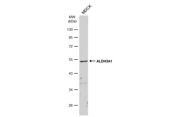
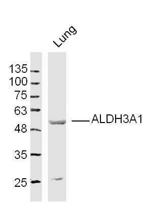
Whole cell extract (30 μg) was separated by 10% SDS-PAGE, and the membrane was blotted with ALDH3A1 antibody (GTX112391) diluted at 1:500. The HRP-conjugated anti-rabbit IgG antibody (GTX213110-01) was used to detect the primary antibody, and the signal was developed with Trident ECL plus-Enhanced.
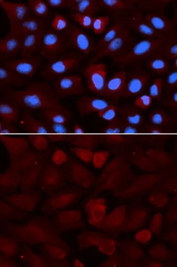
 antibody at 1:100 dilution.
Antigen Retrieval: Trilogy? (EDTA based, pH 8.0) buffer, 15min")
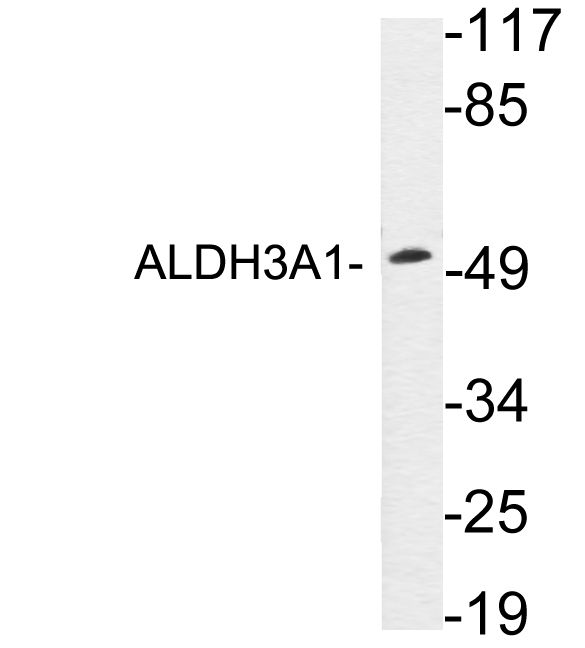
![Whole cell extract (50 μg) was separated by 10% SDS-PAGE, and the membrane was blotted with ALDH3A1 antibody [N1C2] (GTX112391) diluted at 1:500. The HRP-conjugated anti-rabbit IgG antibody (GTX213110-01) was used to detect the primary antibody.](https://www.genetex.com/upload/website/prouct_img/normal/GTX112391/GTX112391_40114_20180817_WB_M_eye_w_23060500_198.webp "Whole cell extract (50 μg) was separated by 10% SDS-PAGE, and the membrane was blotted with ALDH3A1 antibody [N1C2] (GTX112391) diluted at 1:500. The HRP-conjugated anti-rabbit IgG antibody (GTX213110-01) was used to detect the primary antibody.")
Whole cell extract (30 μg) was separated by 10% SDS-PAGE, and the membrane was blotted with ALDH3A1 antibody (GTX112391) diluted at 1:500. The HRP-conjugated anti-rabbit IgG antibody (GTX213110-01) was used to detect the primary antibody, and the signal was developed with Trident ECL plus-Enhanced.
ALDH3A1 antibody [N1C2]
GTX112391
ApplicationsWestern Blot, ImmunoHistoChemistry, ImmunoHistoChemistry Frozen
Product group Antibodies
ReactivityBovine, Canine, Human, Mouse
TargetALDH3A1
Overview
- SupplierGeneTex
- Product NameALDH3A1 antibody [N1C2]
- Delivery Days Customer9
- Application Supplier NoteWB: 1:500-1:3000. IHC-Fr: 1:100-1:1000. *Optimal dilutions/concentrations should be determined by the researcher.Not tested in other applications.
- ApplicationsWestern Blot, ImmunoHistoChemistry, ImmunoHistoChemistry Frozen
- CertificationResearch Use Only
- ClonalityPolyclonal
- Concentration0.61 mg/ml
- ConjugateUnconjugated
- Gene ID218
- Target nameALDH3A1
- Target descriptionaldehyde dehydrogenase 3 family member A1
- Target synonymsALDH3, ALDHIII, aldehyde dehydrogenase, dimeric NADP-preferring, aldehyde dehydrogenase isozyme 3, aldehyde dehydrogenase type III, stomach aldehyde dehydrogenase
- HostRabbit
- IsotypeIgG
- Protein IDP30838
- Protein NameAldehyde dehydrogenase, dimeric NADP-preferring
- Scientific DescriptionAldehyde dehydrogenases oxidize various aldehydes to the corresponding acids. They are involved in the detoxification of alcohol-derived acetaldehyde and in the metabolism of corticosteroids, biogenic amines, neurotransmitters, and lipid peroxidation. The enzyme encoded by this gene forms a cytoplasmic homodimer that preferentially oxidizes aromatic and medium-chain (6 carbons or more) saturated and unsaturated aldehyde substrates. It is thought to promote resistance to UV and 4-hydroxy-2-nonenal-induced oxidative damage in the cornea. The gene is located within the Smith-Magenis syndrome region on chromosome 17. Multiple alternatively spliced variants, encoding the same protein, have been identified. [provided by RefSeq]
- ReactivityBovine, Canine, Human, Mouse
- Storage Instruction-20°C or -80°C,2°C to 8°C
- UNSPSC41116161
Datasheet
Related products
Product group Antibodies
Anti-ALDH3A1 AntibodyA98931
ApplicationsWestern Blot, ELISA
ReactivityHuman, Rat
- SizePrice

Product group Antibodies
Anti-ALDH3A1 Antibody Picoband(r)A01121-3-CARRIER-FREE
ApplicationsImmunoFluorescence, Western Blot, ImmunoCytoChemistry
ReactivityHuman, Mouse, Rat
TargetALDH3A1
- SizePrice
Product group Antibodies
Anti-ALDH3A1 Antibody144-05502
ApplicationsImmunoFluorescence, Western Blot
ReactivityHuman, Mouse
TargetALDH3A1
- SizePrice
Product group Antibodies
ALDH3A1 Polyclonal AntibodyBS-15496R
ApplicationsImmunoFluorescence, Western Blot, ELISA, ImmunoCytoChemistry, ImmunoHistoChemistry, ImmunoHistoChemistry Frozen, ImmunoHistoChemistry Paraffin
ReactivityEquine, Human, Mouse, Porcine, Rat
- SizePrice
Product group Antibodies
ALDH3A1 AntibodyCSB-PA001572EA01HU
ApplicationsImmunoFluorescence, Western Blot, ELISA, ImmunoHistoChemistry
ReactivityHuman
TargetALDH3A1
- SizePrice
Product group Antibodies

Goat anti-ALDH3A1EB10119
ApplicationsWestern Blot, ELISA, ImmunoHistoChemistry
ReactivityHuman, Mouse
TargetALDH3A1
- SizePrice
Product group Antibodies
Aldh3A1 Polyclonal AntibodyCAC07162
ApplicationsImmunoFluorescence, Western Blot, ELISA, ImmunoHistoChemistry
TargetALDH3A1
- SizePrice
Product group Antibodies

ALDH3A1 AntibodyLS-C404481
ApplicationsWestern Blot, ELISA, ImmunoHistoChemistry
ReactivityHuman, Mouse, Rat
TargetALDH3A1
- SizePrice
Product group Antibodies
References
ALDH3A1 antibodyGTX30042
ApplicationsImmunoFluorescence, Western Blot, ImmunoCytoChemistry
ReactivityHuman, Mouse
TargetALDH3A1
- SizePrice