

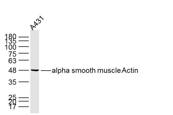
alpha Smooth Muscle Actin antibody [0.N.5]
GTX18147
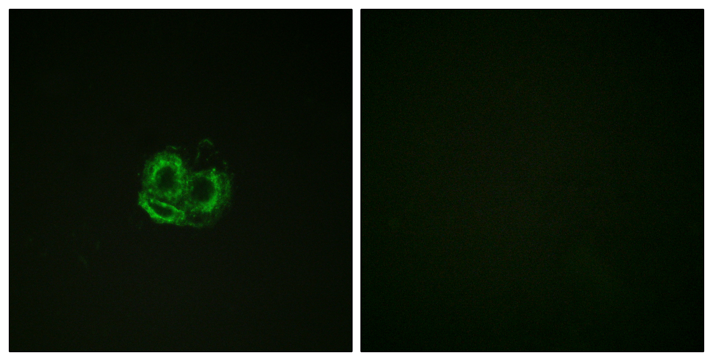
ApplicationsImmunoFluorescence, ImmunoCytoChemistry, ImmunoHistoChemistry, ImmunoHistoChemistry Paraffin
Product group Antibodies
ReactivityHuman, Mouse, Rabbit
TargetACTA2
Overview
- SupplierGeneTex
- Product Namealpha Smooth Muscle Actin antibody [0.N.5]
- Delivery Days Customer9
- Application Supplier NoteIHC-P: 1:800 for 20 minutes at RT. *Optimal dilutions/concentrations should be determined by the researcher.Not tested in other applications.
- ApplicationsImmunoFluorescence, ImmunoCytoChemistry, ImmunoHistoChemistry, ImmunoHistoChemistry Paraffin
- CertificationResearch Use Only
- ClonalityMonoclonal
- Clone ID0.N.5
- ConjugateUnconjugated
- Gene ID59
- Target nameACTA2
- Target descriptionactin alpha 2, smooth muscle
- Target synonymsACTSA, SMDYS, actin, aortic smooth muscle, actin, alpha 2, smooth muscle, aorta, alpha-cardiac actin, cell growth-inhibiting gene 46 protein
- HostMouse
- IsotypeIgG2a
- Protein IDP62736
- Protein NameActin, aortic smooth muscle
- Scientific DescriptionThis gene encodes one of six different actin proteins. Actins are highly conserved proteins that are involved in cell motility, structure, integrity, and intercellular signaling. The encoded protein is a smooth muscle actin that is involved in vascular contractility and blood pressure homeostasis. Mutations in this gene cause a variety of vascular diseases, such as thoracic aortic disease, coronary artery disease, stroke, and Moyamoya disease, as well as multisystemic smooth muscle dysfunction syndrome. [provided by RefSeq, Sep 2017]
- ReactivityHuman, Mouse, Rabbit
- Storage Instruction-20°C or -80°C,2°C to 8°C
- UNSPSC12352203
References
- Xie X, Wu Q, Liu Y, et al. Vascular endothelial growth factor attenuates neointimal hyperplasia of decellularized small-diameter vascular grafts by modulating the local inflammatory response. Front Bioeng Biotechnol. 2022,10:1066266. doi: 10.3389/fbioe.2022.1066266Read this paper
- Chen C, Lu T, Wu Z, et al. A proteomics analysis of neointima formation on decellularized vascular grafts reveals regenerative alterations in protein signature running head: Proteomics analysis of neointima formation. Front Bioeng Biotechnol. 2022,10:894956. doi: 10.3389/fbioe.2022.894956Read this paper
- Taghizadeh H, Taghizadehghalehjoughi A, Yildirim S, et al. Deteriorated Vascular Homeostasis in Hypertension: Experimental Evidence from Aorta, Brain, and Pancreatic Vasculature. J Pers Med. 2022,12(10). doi: 10.3390/jpm12101602Read this paper
- Liu Y, Chen C, Xie X, et al. Photooxidation and Pentagalloyl Glucose Cross-Linking Improves the Performance of Decellularized Small-Diameter Vascular Xenograft In Vivo. Front Bioeng Biotechnol. 2022,10:816513. doi: 10.3389/fbioe.2022.816513Read this paper
- Riggle BA, Manglani M, Maric D, et al. CD8+ T cells target cerebrovasculature in children with cerebral malaria. J Clin Invest. 2020,130(3):1128-1138. doi: 10.1172/JCI133474Read this paper
- Otto C, Schkoldow J, Krahl E, et al. Use of a murine endometriosis interna model for the characterization of compounds that effectively treat human endometriosis. Exp Ther Med. 2012,3(3):410-414.Read this paper
- Klarquist J, Barfuss A, Kandala S, et al. Melanoma-associated antigen expression in lymphangioleiomyomatosis renders tumor cells susceptible to cytotoxic T cells. Am J Pathol. 2009,175(6):2463-72. doi: 10.2353/ajpath.2009.090525Read this paper
Datasheet
Related products
Product group Antibodies

Anti-alpha-SMA [8E5D12A9]Ab03397-1.1
ApplicationsWestern Blot, ELISA, ImmunoHistoChemistry
ReactivityHuman
TargetACTA2
- SizePrice
Product group Antibodies
Anti-ACTA2 Antibody144-07248
ApplicationsImmunoFluorescence, Western Blot
ReactivityHuman, Mouse, Rat
TargetACTA2
- SizePrice
Product group Antibodies
References
ApplicationsFlow Cytometry, ImmunoFluorescence, Western Blot, ImmunoHistoChemistry, ImmunoHistoChemistry Frozen, ImmunoHistoChemistry Paraffin
ReactivityHuman, Mouse, Rat
TargetACTA2
- SizePrice
Product group Antibodies
ACTA2 Monoclonal AntibodyCSB-MA080158
ApplicationsWestern Blot, ELISA, ImmunoHistoChemistry
ReactivityHuman, Mouse, Rat
TargetACTA2
- SizePrice
Product group Antibodies
References
ApplicationsImmunoFluorescence, Western Blot, ImmunoCytoChemistry, ImmunoHistoChemistry, ImmunoHistoChemistry Frozen, ImmunoHistoChemistry Paraffin
ReactivityHuman, Mouse, Rat
TargetACTA2
- SizePrice
Product group Antibodies
anti-ACTA2, Rabbit Monoclonal (RM253)REV-31-1133-00
ApplicationsWestern Blot, ImmunoHistoChemistry
ReactivityHuman, Mouse
TargetACTA2
- SizePrice
Product group Antibodies
Anti-Actin-pan AntibodyA94548
ApplicationsImmunoFluorescence, Western Blot, ELISA, ImmunoHistoChemistry
ReactivityHuman, Mouse, Rat
- SizePrice
![WB analysis of various sample lysates using GTX34111 alpha Smooth Muscle Actin antibody [6A12]. Lane 1 : HeLa cell lysate Lane 2 : 3T3 cell lysate Lane 3 : Rat brain tissue lysate](https://www.genetex.com/upload/website/prouct_img/normal/GTX34111/GTX34111_20200622_WB_522_w_23060801_487.webp)
Product group Antibodies
ApplicationsWestern Blot, ImmunoHistoChemistry, ImmunoHistoChemistry Paraffin
ReactivityHuman, Mouse, Rat
TargetACTA2
- SizePrice
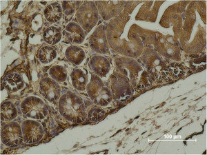
![IHC-P analysis of human small bowel tissue using GTX44499 alpha Smooth Muscle Actin antibody [αsm-1]. Note cytoplasmic staining of the muscularis mucosa, vascular walls and smooth muscle fibers in the lamina propria.](https://www.genetex.com/upload/website/prouct_img/normal/GTX44499/GTX44499_20200811_IHC-P_5_w_23060820_700.webp)
Product group Antibodies
ApplicationsImmunoHistoChemistry, ImmunoHistoChemistry Paraffin
ReactivityHuman
TargetACTA2
- SizePrice