

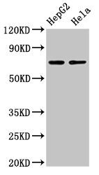
Western Blot Positive WB detected in: HepG2 whole cell lysate, Hela whole cell lysate All lanes: AMFR antibody at 4microg/ml Secondary Goat polyclonal to rabbit IgG at 1/50000 dilution Predicted band size: 73 kDa Observed band size: 73 kDa
. Section was blocked with 10% normal goat serum 30min at RT. Then primary antibody (1% BSA) was incubated at 4°C overnight. The primary is detected by a biotinylated secondary antibody and visualized using an HRP conjugated SP system.")
. Section was blocked with 10% normal goat serum 30min at RT. Then primary antibody (1% BSA) was incubated at 4°C overnight. The primary is detected by a biotinylated secondary antibody and visualized using an HRP conjugated SP system.")
Western Blot Positive WB detected in: HepG2 whole cell lysate, Hela whole cell lysate All lanes: AMFR antibody at 4microg/ml Secondary Goat polyclonal to rabbit IgG at 1/50000 dilution Predicted band size: 73 kDa Observed band size: 73 kDa
AMFR Antibody
CSB-PA333164LA01HU
ApplicationsWestern Blot, ELISA, ImmunoHistoChemistry
Product group Antibodies
ReactivityHuman
TargetAMFR
Overview
- SupplierCusabio
- Product NameAMFR Antibody
- Delivery Days Customer20
- ApplicationsWestern Blot, ELISA, ImmunoHistoChemistry
- CertificationResearch Use Only
- ClonalityPolyclonal
- ConjugateUnconjugated
- Gene ID267
- Target nameAMFR
- Target descriptionautocrine motility factor receptor
- Target synonymsGP78, RNF45, SPG89, E3 ubiquitin-protein ligase AMFR, RING finger protein 45, RING-type E3 ubiquitin transferase AMFR
- HostRabbit
- IsotypeIgG
- Protein IDQ9UKV5
- Protein NameE3 ubiquitin-protein ligase AMFR
- Scientific DescriptionE3 ubiquitin-protein ligase that mediates the polyubiquitination of a number of proteins such as CD3D, CYP3A4, CFTR and APOB for proteasomal degradation. Component of a VCP/p97-AMFR/gp78 complex that participates in the final step of endoplasmic reticulum-associated degradation (ERAD). The VCP/p97-AMFR/gp78 complex is involved in the sterol-accelerated ERAD degradation of HMGCR through binding to the HMGCR-INSIG complex at the ER membrane and initiating ubiquitination of HMGCR. The ubiquitinated HMGCR is then released from the ER by the complex into the cytosol for subsequent destruction. Also regulates ERAD through the ubiquitination of UBL4A a component of the BAG6/BAT3 complex. Also acts as a scaffold protein to assemble a complex that couples ubiquitination, retranslocation and deglycosylation. Mediates tumor invasion and metastasis as a receptor for the GPI/autocrine motility factor.
- ReactivityHuman
- Storage Instruction-20°C or -80°C
- UNSPSC41116161
Related products

Product group Antibodies
Anti-AMFR AntibodyHPA029018
ApplicationsImmunoHistoChemistry
ReactivityHuman
TargetAMFR
- SizePrice
Product group Antibodies

GP78 / AMFR AntibodyLS-C669761
ApplicationsWestern Blot, ELISA, ImmunoHistoChemistry, ImmunoHistoChemistry Paraffin
ReactivityHuman
TargetAMFR
- SizePrice
Product group Antibodies
AMFR Polyclonal AntibodyCAC15127
ApplicationsWestern Blot, ELISA, ImmunoHistoChemistry
TargetAMFR
- SizePrice

Product group Antibodies
Anti-AMFR Antibody Picoband(r)PB10039-CARRIER-FREE
ApplicationsFlow Cytometry, ImmunoFluorescence, Western Blot, ImmunoCytoChemistry, ImmunoHistoChemistry
ReactivityHuman, Mouse, Rat
TargetAMFR
- SizePrice
![Boiled and unboiled mouse tissue extract (50 μg) were separated by 7.5% SDS-PAGE, and the membrane was blotted with AMFR antibody [HL2865] (GTX640142) diluted at 1:1000. The HRP-conjugated anti-rabbit IgG antibody (GTX213110-01) was used to detect the primary antibody.](https://www.genetex.com/upload/website/prouct_img/normal/GTX640142/GTX640142_T-45362_20240412_WB_M_liver_24041523_454.webp)
Product group Antibodies
AMFR antibody [HL2865]GTX640142
ApplicationsWestern Blot, ImmunoHistoChemistry, ImmunoHistoChemistry Paraffin
ReactivityHuman, Mouse, Rat
TargetAMFR
- SizePrice
Product group Antibodies
Anti-AMFR Antibody144-65732
ApplicationsWestern Blot
ReactivityHuman
TargetAMFR
- SizePrice
Product group Antibodies
RNF45 Polyclonal AntibodyBS-6511R
ApplicationsImmunoFluorescence, Western Blot, ELISA, ImmunoCytoChemistry, ImmunoHistoChemistry, ImmunoHistoChemistry Frozen, ImmunoHistoChemistry Paraffin
ReactivityBovine, Canine, Chicken, Human, Mouse, Rabbit, Rat
TargetAMFR
- SizePrice