

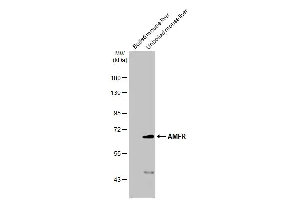
Boiled and unboiled mouse tissue extract (50 μg) were separated by 7.5% SDS-PAGE, and the membrane was blotted with AMFR antibody [HL2865] (GTX640142) diluted at 1:1000. The HRP-conjugated anti-rabbit IgG antibody (GTX213110-01) was used to detect the primary antibody.
![Boiled and unboiled rat tissue extract (50 μg) were separated by 7.5% SDS-PAGE, and the membrane was blotted with AMFR antibody [HL2865] (GTX640142) diluted at 1:1000. The HRP-conjugated anti-rabbit IgG antibody (GTX213110-01) was used to detect the primary antibody.](https://www.genetex.com/upload/website/prouct_img/normal/GTX640142/GTX640142_T-45362_20240412_WB_R_liver_24041523_650.webp "Boiled and unboiled rat tissue extract (50 μg) were separated by 7.5% SDS-PAGE, and the membrane was blotted with AMFR antibody [HL2865] (GTX640142) diluted at 1:1000. The HRP-conjugated anti-rabbit IgG antibody (GTX213110-01) was used to detect the primary antibody.")
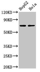
![Boiled and unboiled HepG2 whole cell and membrane extracts (30 μg) were separated by 7.5% SDS-PAGE, and the membrane was blotted with AMFR antibody [HL2865] (GTX640142) diluted at 1:1000. The HRP-conjugated anti-rabbit IgG antibody (GTX213110-01) was used to detect the primary antibody. (WCE: whole cell extract; ME: membrane extract)](https://www.genetex.com/upload/website/prouct_img/normal/GTX640142/GTX640142_45425_20240607_WB_Fraction_24061301_154.webp "Boiled and unboiled HepG2 whole cell and membrane extracts (30 μg) were separated by 7.5% SDS-PAGE, and the membrane was blotted with AMFR antibody [HL2865] (GTX640142) diluted at 1:1000. The HRP-conjugated anti-rabbit IgG antibody (GTX213110-01) was used to detect the primary antibody. (WCE: whole cell extract; ME: membrane extract)")
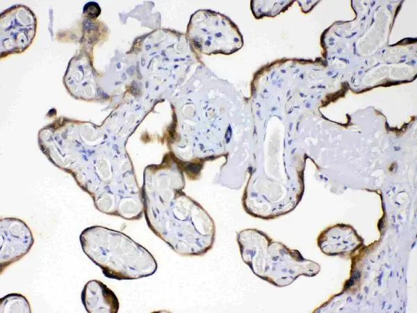
![AMFR antibody [HL2865] detects AMFR protein by immunohistochemical analysis. Sample: Paraffin-embedded mouse liver. AMFR stained by AMFR antibody [HL2865] (GTX640142) diluted at 1:100. Antigen Retrieval: Citrate buffer, pH 6.0, 20 min](https://www.genetex.com/upload/website/prouct_img/normal/GTX640142/GTX640142_T-45362_20240719_IHC-P_M_24080622_524.webp "AMFR antibody [HL2865] detects AMFR protein by immunohistochemical analysis. Sample: Paraffin-embedded mouse liver. AMFR stained by AMFR antibody [HL2865] (GTX640142) diluted at 1:100. Antigen Retrieval: Citrate buffer, pH 6.0, 20 min")
Boiled and unboiled mouse tissue extract (50 μg) were separated by 7.5% SDS-PAGE, and the membrane was blotted with AMFR antibody [HL2865] (GTX640142) diluted at 1:1000. The HRP-conjugated anti-rabbit IgG antibody (GTX213110-01) was used to detect the primary antibody.
AMFR antibody [HL2865]
GTX640142
ApplicationsWestern Blot, ImmunoHistoChemistry, ImmunoHistoChemistry Paraffin
Product group Antibodies
ReactivityHuman, Mouse, Rat
TargetAMFR
Overview
- SupplierGeneTex
- Product NameAMFR antibody [HL2865]
- Delivery Days Customer7
- Application Supplier NoteWB: 1:500-1:3000. *Optimal dilutions/concentrations should be determined by the researcher.Not tested in other applications.
- ApplicationsWestern Blot, ImmunoHistoChemistry, ImmunoHistoChemistry Paraffin
- CertificationResearch Use Only
- ClonalityMonoclonal
- Clone IDHL2865
- Concentration1 mg/ml
- ConjugateUnconjugated
- Gene ID267
- Target nameAMFR
- Target descriptionautocrine motility factor receptor
- Target synonymsGP78, RNF45, SPG89, E3 ubiquitin-protein ligase AMFR, RING finger protein 45, RING-type E3 ubiquitin transferase AMFR
- HostRabbit
- IsotypeIgG
- Protein IDQ9UKV5
- Protein NameE3 ubiquitin-protein ligase AMFR
- Scientific DescriptionThis locus encodes a glycosylated transmembrane receptor. Its ligand, autocrine motility factor, is a tumor motility-stimulating protein secreted by tumor cells. The encoded receptor is also a member of the E3 ubiquitin ligase family of proteins. It catalyzes ubiquitination and endoplasmic reticulum-associated degradation of specific proteins. [provided by RefSeq, Feb 2012]
- ReactivityHuman, Mouse, Rat
- Storage Instruction-20°C or -80°C,2°C to 8°C
- UNSPSC41116161
Datasheet
Related products
Product group Antibodies
AMFR AntibodyCSB-PA333164LA01HU
ApplicationsWestern Blot, ELISA, ImmunoHistoChemistry
ReactivityHuman
TargetAMFR
- SizePrice

Product group Antibodies
Anti-AMFR AntibodyHPA029018
ApplicationsImmunoHistoChemistry
ReactivityHuman
TargetAMFR
- SizePrice
Product group Antibodies

GP78 / AMFR AntibodyLS-C669761
ApplicationsWestern Blot, ELISA, ImmunoHistoChemistry, ImmunoHistoChemistry Paraffin
ReactivityHuman
TargetAMFR
- SizePrice
Product group Antibodies
AMFR Polyclonal AntibodyCAC15127
ApplicationsWestern Blot, ELISA, ImmunoHistoChemistry
TargetAMFR
- SizePrice

Product group Antibodies
Anti-AMFR Antibody Picoband(r)PB10039-CARRIER-FREE
ApplicationsFlow Cytometry, ImmunoFluorescence, Western Blot, ImmunoCytoChemistry, ImmunoHistoChemistry
ReactivityHuman, Mouse, Rat
TargetAMFR
- SizePrice
![Boiled and unboiled mouse tissue extract (50 μg) were separated by 7.5% SDS-PAGE, and the membrane was blotted with AMFR antibody [HL2866] (GTX640143) diluted at 1:1000. The HRP-conjugated anti-rabbit IgG antibody (GTX213110-01) was used to detect the primary antibody.](https://www.genetex.com/upload/website/prouct_img/normal/GTX640143/GTX640143_T-45362_20240607_WB_M_liver_24061301_878.webp)
Product group Antibodies
AMFR antibody [HL2866]GTX640143
ApplicationsImmunoFluorescence, Western Blot, ImmunoCytoChemistry
ReactivityHuman, Mouse
TargetAMFR
- SizePrice
Product group Antibodies
AMFR antibodyGTX04315
ApplicationsFlow Cytometry, ImmunoFluorescence, Western Blot, ImmunoCytoChemistry, ImmunoHistoChemistry, ImmunoHistoChemistry Paraffin
ReactivityHuman, Mouse, Rat
TargetAMFR
- SizePrice
Product group Antibodies
Anti-AMFR Antibody144-65732
ApplicationsWestern Blot
ReactivityHuman
TargetAMFR
- SizePrice
Product group Antibodies
RNF45 Polyclonal AntibodyBS-6511R
ApplicationsImmunoFluorescence, Western Blot, ELISA, ImmunoCytoChemistry, ImmunoHistoChemistry, ImmunoHistoChemistry Frozen, ImmunoHistoChemistry Paraffin
ReactivityBovine, Canine, Chicken, Human, Mouse, Rabbit, Rat
TargetAMFR
- SizePrice