

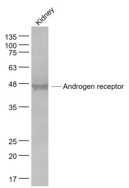
WB analysis of mouse kidney tissue lysates using GTX03929 Androgen Receptor antibody. Dilution : 1:1000
 15 min Dilution : 1:500")

WB analysis of mouse kidney tissue lysates using GTX03929 Androgen Receptor antibody. Dilution : 1:1000
Androgen Receptor antibody
GTX03929
ApplicationsImmunoFluorescence, Western Blot, ImmunoCytoChemistry, ImmunoHistoChemistry, ImmunoHistoChemistry Paraffin
Product group Antibodies
ReactivityGoat, Human, Mouse, Porcine, Rat
TargetAR
Overview
- SupplierGeneTex
- Product NameAndrogen Receptor antibody
- Delivery Days Customer9
- Application Supplier NoteWB: 1:300-1:5000. ICC/IF: 1:50-1:200. IHC-P: 1:200-1:400. *Optimal dilutions/concentrations should be determined by the researcher.Not tested in other applications.
- ApplicationsImmunoFluorescence, Western Blot, ImmunoCytoChemistry, ImmunoHistoChemistry, ImmunoHistoChemistry Paraffin
- CertificationResearch Use Only
- ClonalityPolyclonal
- Concentration1 mg/ml
- ConjugateUnconjugated
- Gene ID367
- Target nameAR
- Target descriptionandrogen receptor
- Target synonymsAIS, AR8, DHTR, HUMARA, HYSP1, KD, NR3C4, SBMA, SMAX1, TFM, androgen receptor, dihydrotestosterone receptor, nuclear receptor subfamily 3 group C member 4
- HostRabbit
- IsotypeIgG
- Protein IDP10275
- Protein NameAndrogen receptor
- Scientific DescriptionThe androgen receptor gene is more than 90 kb long and codes for a protein that has 3 major functional domains: the N-terminal domain, DNA-binding domain, and androgen-binding domain. The protein functions as a steroid-hormone activated transcription factor. Upon binding the hormone ligand, the receptor dissociates from accessory proteins, translocates into the nucleus, dimerizes, and then stimulates transcription of androgen responsive genes. This gene contains 2 polymorphic trinucleotide repeat segments that encode polyglutamine and polyglycine tracts in the N-terminal transactivation domain of its protein. Expansion of the polyglutamine tract from the normal 9-34 repeats to the pathogenic 38-62 repeats causes spinal bulbar muscular atrophy (SBMA, also known as Kennedys disease). Mutations in this gene are also associated with complete androgen insensitivity (CAIS). Alternative splicing results in multiple transcript variants encoding different isoforms. [provided by RefSeq, Jan 2017]
- ReactivityGoat, Human, Mouse, Porcine, Rat
- Storage Instruction-20°C or -80°C,2°C to 8°C
- UNSPSC12352203
Datasheet
Related products
Product group Antibodies

ApplicationsWestern Blot, ELISA
- SizePrice
Product group Antibodies
Anti-Androgen Receptor Antibody130-10000
ApplicationsWestern Blot, ELISA
ReactivityHuman
TargetAR
- SizePrice

Product group Antibodies
Anti-Androgen Receptor/AR Antibody Picoband(r)A00542-CARRIER-FREE
ApplicationsFlow Cytometry, ImmunoFluorescence, Western Blot, ELISA, ImmunoCytoChemistry
ReactivityHuman, Mouse, Rat
TargetAR
- SizePrice
![WB analysis of mouse testis tissue lysate using GTX01536 Androgen Receptor antibody [GT1228]. Dilution : 1:1000 Loading : 25 μg](https://www.genetex.com/upload/website/prouct_img/normal/GTX01536/GTX01536_20200508_WB_w_23053121_665.webp)
Product group Antibodies
ApplicationsImmunoFluorescence, Western Blot, ImmunoCytoChemistry, ImmunoHistoChemistry, ImmunoHistoChemistry Paraffin
ReactivityHuman, Mouse
TargetAR
- SizePrice
![IHC-P analysis of human prostate tissue using GTX04368 Androgen Receptor antibody [MSVA-367R] HistoMAX?. A strong nuclear androgen receptor immunostaining is seen in stromal and epithelial cells of the prostate.](https://www.genetex.com/upload/website/prouct_img/normal/GTX04368/GTX04368_20230728_IHC-P_3_23072722_392.webp)
Product group Antibodies
ApplicationsImmunoHistoChemistry, ImmunoHistoChemistry Paraffin
ReactivityHuman
TargetAR
- SizePrice
![Various whole cell extracts (30 μg) were separated by 5% SDS-PAGE, and the membrane was blotted with Androgen Receptor antibody [N1], N-term (GTX100056) diluted at 1:500. The HRP-conjugated anti-rabbit IgG antibody (GTX213110-01) was used to detect the primary antibody.](https://www.genetex.com/upload/website/prouct_img/normal/GTX100056/GTX100056_44070_20201008_WB_22092119_792.webp)
Product group Antibodies
References
ApplicationsImmunoFluorescence, Western Blot, ImmunoCytoChemistry, ImmunoHistoChemistry, ImmunoHistoChemistry Paraffin
ReactivityHuman, Mouse
TargetAR
- SizePrice
![Androgen Receptor (ARv7 Splice Variant) antiboy [HL1028] detects Androgen Receptor (ARv7 Splice Variant) protein at nucleus by immunofluorescent analysis. Sample: 22RV1 cells were fixed in 4% paraformaldehyde at RT for 15 min. Green: Androgen Receptor (ARv7 Splice Variant) stained by Androgen Receptor (ARv7 Splice Variant) antiboy [HL1028] (GTX635842) diluted at 1:500. Red: alpha Tubulin, a cytoskeleton marker, stained by alpha Tubulin antibody [GT114] (GTX628802) diluted at 1:1000.](https://www.genetex.com/upload/website/prouct_img/normal/GTX635842/GTX635842_T-44130_20211126_ICC_IF_w_23061202_224.webp)
Product group Antibodies
ApplicationsImmunoFluorescence, Western Blot, ImmunoCytoChemistry, ImmunoHistoChemistry, ImmunoHistoChemistry Paraffin
ReactivityHuman
TargetAR
- SizePrice
![Androgen Receptor antibody [HL1049] detects Androgen Receptor protein by immunohistochemical analysis. Sample: Paraffin-embedded rat tissues. Androgen Receptor stained by Androgen Receptor antibody [HL1049] (GTX636021) diluted at 1:100. Antigen Retrieval: Citrate buffer, pH 6.0, 15 min](https://www.genetex.com/upload/website/prouct_img/normal/GTX636021/GTX636021_44277_20221223_IHC-P_multiple_R_22122821_517.webp)
Product group Antibodies
Androgen Receptor antibody [HL1049]GTX636021
ApplicationsImmunoFluorescence, Western Blot, ImmunoCytoChemistry, ImmunoHistoChemistry, ImmunoHistoChemistry Paraffin
ReactivityHuman, Rat
TargetAR
- SizePrice
![Androgen Receptor (ARv7 Splice Variant) antibody [HL1239] detects Androgen Receptor (ARv7 Splice Variant) protein at nucleus by immunohistochemical analysis. Sample: PC-3 and 22RV1 FFPE Cell Pellet Block. Green: Androgen Receptor (ARv7 Splice Variant) stained by Androgen Receptor (ARv7 Splice Variant) antibody [HL1239] (GTX636638) diluted at 1:200. Blue: Fluoroshield with DAPI (GTX30920). Antigen Retrieval: Citrate buffer, pH 6.0, 15 min](https://www.genetex.com/upload/website/prouct_img/normal/GTX636638/GTX636638_44578_20220318_IHC-P_cell_pellet_w_23061202_242.webp)
Product group Antibodies
ApplicationsImmunoFluorescence, Western Blot, ImmunoCytoChemistry, ImmunoHistoChemistry
ReactivityHuman
TargetAR
- SizePrice