

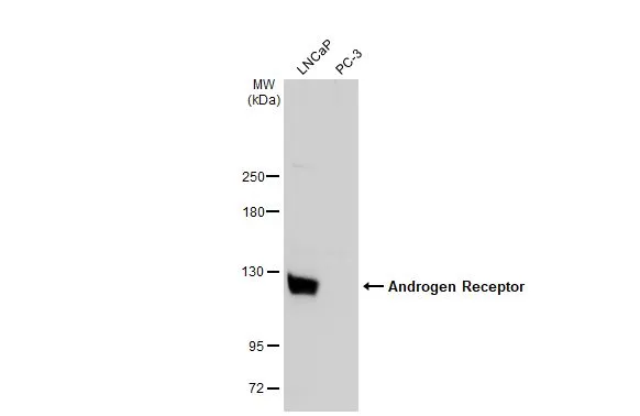
Various whole cell extracts (30 μg) were separated by 5% SDS-PAGE, and the membrane was blotted with Androgen Receptor antibody [N1], N-term (GTX100056) diluted at 1:500. The HRP-conjugated anti-rabbit IgG antibody (GTX213110-01) was used to detect the primary antibody.
![Androgen Receptor antibody [N1], N-term detects Androgen Receptor protein at nucleus by immunofluorescent analysis. Sample: LNCap cells were fixed in 4% paraformaldehyde at RT for 15 min. Green: Androgen Receptor stained by Androgen Receptor antibody [N1], N-term (GTX100056) diluted at 1:2000.](https://www.genetex.com/upload/website/prouct_img/normal/GTX100056/GTX100056_44069_20210122_ICC_IF_w_23053123_464.webp "Androgen Receptor antibody [N1], N-term detects Androgen Receptor protein at nucleus by immunofluorescent analysis. Sample: LNCap cells were fixed in 4% paraformaldehyde at RT for 15 min. Green: Androgen Receptor stained by Androgen Receptor antibody [N1], N-term (GTX100056) diluted at 1:2000.")
![Androgen Receptor antibody [N1], N-term detects Androgen Receptor protein at cytoplasm and nucleus by immunohistochemical analysis. Sample: Paraffin-embedded mouse testis. Androgen Receptor stained by Androgen Receptor antibody [N1], N-term (GTX100056) diluted at 1:500. Antigen Retrieval: Citrate buffer, pH 6.0, 15 min](https://www.genetex.com/upload/website/prouct_img/normal/GTX100056/GTX100056_44069_20201023_IHC-P_M_w_23053123_594.webp "Androgen Receptor antibody [N1], N-term detects Androgen Receptor protein at cytoplasm and nucleus by immunohistochemical analysis. Sample: Paraffin-embedded mouse testis. Androgen Receptor stained by Androgen Receptor antibody [N1], N-term (GTX100056) diluted at 1:500. Antigen Retrieval: Citrate buffer, pH 6.0, 15 min")
![Androgen Receptor antibody [N1], N-term detects Androgen Receptor protein at nucleus on mouse prostate by immunohistochemical analysis. Sample: Paraffin-embedded mouse prostate. Androgen Receptor antibody [N1], N-term (GTX100056) dilution: 1:500.
Antigen Retrieval: Trilogy? (EDTA based, pH 8.0) buffer, 15min](https://www.genetex.com/upload/website/prouct_img/normal/GTX100056/GTX100056_39476_IHC_M_w_23053123_269.webp "Androgen Receptor antibody [N1], N-term detects Androgen Receptor protein at nucleus on mouse prostate by immunohistochemical analysis. Sample: Paraffin-embedded mouse prostate. Androgen Receptor antibody [N1], N-term (GTX100056) dilution: 1:500.
Antigen Retrieval: Trilogy? (EDTA based, pH 8.0) buffer, 15min")

Various whole cell extracts (30 μg) were separated by 5% SDS-PAGE, and the membrane was blotted with Androgen Receptor antibody [N1], N-term (GTX100056) diluted at 1:500. The HRP-conjugated anti-rabbit IgG antibody (GTX213110-01) was used to detect the primary antibody.
Androgen Receptor antibody [N1], N-term
GTX100056
ApplicationsImmunoFluorescence, Western Blot, ImmunoCytoChemistry, ImmunoHistoChemistry, ImmunoHistoChemistry Paraffin
Product group Antibodies
ReactivityHuman, Mouse
TargetAR
Overview
- SupplierGeneTex
- Product NameAndrogen Receptor antibody [N1], N-term
- Delivery Days Customer9
- Application Supplier NoteWB: 1:500-1:3000. ICC/IF: 1:100-1:1000. IHC-P: 1:100-1:1000. *Optimal dilutions/concentrations should be determined by the researcher.Not tested in other applications.
- ApplicationsImmunoFluorescence, Western Blot, ImmunoCytoChemistry, ImmunoHistoChemistry, ImmunoHistoChemistry Paraffin
- CertificationResearch Use Only
- ClonalityPolyclonal
- Concentration0.47 mg/ml
- ConjugateUnconjugated
- Gene ID367
- Target nameAR
- Target descriptionandrogen receptor
- Target synonymsAIS, AR8, DHTR, HUMARA, HYSP1, KD, NR3C4, SBMA, SMAX1, TFM, androgen receptor, dihydrotestosterone receptor, nuclear receptor subfamily 3 group C member 4
- HostRabbit
- IsotypeIgG
- Protein IDP10275
- Protein NameAndrogen receptor
- Scientific DescriptionThe androgen receptor gene is more than 90 kb long and codes for a protein that has 3 major functional domains: the N-terminal domain, DNA-binding domain, and androgen-binding domain. The protein functions as a steroid-hormone activated transcription factor. Upon binding the hormone ligand, the receptor dissociates from accessory proteins, translocates into the nucleus, dimerizes, and then stimulates transcription of androgen responsive genes. This gene contains 2 polymorphic trinucleotide repeat segments that encode polyglutamine and polyglycine tracts in the N-terminal transactivation domain of its protein. Expansion of the polyglutamine tract causes spinal bulbar muscular atrophy (Kennedy disease). Mutations in this gene are also associated with complete androgen insensitivity (CAIS). Two alternatively spliced variants encoding distinct isoforms have been described. [provided by RefSeq]
- ReactivityHuman, Mouse
- Storage Instruction-20°C or -80°C,2°C to 8°C
- UNSPSC41116161
Datasheet
Related products
Product group Antibodies
AR AntibodyCSB-PA001975XA01HU
ApplicationsWestern Blot, ELISA
ReactivityHuman
TargetAR
- SizePrice

Product group Antibodies
Anti-Androgen Receptor/AR Antibody Picoband(r)A00542-CARRIER-FREE
ApplicationsFlow Cytometry, ImmunoFluorescence, Western Blot, ELISA, ImmunoCytoChemistry
ReactivityHuman, Mouse, Rat
TargetAR
- SizePrice
Product group Antibodies
ApplicationsWestern Blot, ELISA
ReactivityHuman, Mouse, Rat
- SizePrice
Product group Antibodies

ApplicationsWestern Blot, ELISA
- SizePrice

Product group Antibodies
Anti-AR AntibodyAMAB91547
ApplicationsWestern Blot, ImmunoHistoChemistry
ReactivityHuman
TargetAR
- SizePrice
Product group Antibodies

ApplicationsWestern Blot, ELISA
ReactivityHuman, Mouse, Rat
TargetAR
- SizePrice
Product group Antibodies

Goat anti-Androgen ReceptorEB06441
ApplicationsImmunoFluorescence, Western Blot, ChIP Chromatin ImmunoPrecipitation, ELISA, ImmunoHistoChemistry
ReactivityBovine, Canine, Human, Mouse, Porcine, Rat
TargetAR
- SizePrice
![IHC-P analysis of human prostate carcinoma tissue using GTX29474 Androgen Receptor antibody [AR 441].](https://www.genetex.com/upload/website/prouct_img/normal/GTX29474/GTX29474_20191203_IHC-P_132_w_23060722_648.webp)
Product group Antibodies
References
ApplicationsImmunoHistoChemistry, ImmunoHistoChemistry Paraffin
ReactivityHuman
TargetAR
- SizePrice