

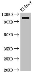
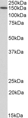
Western Blot Positive WB detected in: Mouse kidney tissue All lanes: ANPEP antibody at 3.4microg/ml Secondary Goat polyclonal to rabbit IgG at 1/50000 dilution Predicted band size: 110 kDa Observed band size: 110 kDa


")
Western Blot Positive WB detected in: Mouse kidney tissue All lanes: ANPEP antibody at 3.4microg/ml Secondary Goat polyclonal to rabbit IgG at 1/50000 dilution Predicted band size: 110 kDa Observed band size: 110 kDa
ANPEP Antibody
CSB-PA001827LA01HU
ApplicationsImmunoFluorescence, Western Blot, ELISA, ImmunoHistoChemistry
Product group Antibodies
ReactivityHuman, Mouse
TargetANPEP
Overview
- SupplierCusabio
- Product NameANPEP Antibody
- Delivery Days Customer20
- ApplicationsImmunoFluorescence, Western Blot, ELISA, ImmunoHistoChemistry
- CertificationResearch Use Only
- ClonalityPolyclonal
- ConjugateUnconjugated
- Gene ID290
- Target nameANPEP
- Target descriptionalanyl aminopeptidase, membrane
- Target synonymsAP-M, AP-N, APN, CD13, GP150, LAP1, P150, PEPN, hAPN, aminopeptidase N, alanyl (membrane) aminopeptidase, aminopeptidase M, membrane alanyl aminopeptidase, microsomal aminopeptidase, myeloid plasma membrane glycoprotein CD13
- HostRabbit
- IsotypeIgG
- Protein IDP15144
- Protein NameAminopeptidase N
- Scientific DescriptionBroad specificity aminopeptidase. Plays a role in the final digestion of peptides generated from hydrolysis of proteins by gastric and pancreatic proteases. May play a critical role in the pathogenesis of cholesterol gallstone disease. May be involved in the metabolism of regulatory peptides of diverse cell types, responsible for the processing of peptide hormones, such as angiotensin III and IV, neuropeptides, and chemokines. Found to cleave antigen peptides bound to major histocompatibility complex class II molecules of presenting cells and to degrade neurotransmitters at synaptic junctions. Is also implicated as a regulator of IL-8 bioavailability in the endometrium, and therefore may contribute to the regulation of angiogenesis. Is used as a marker for acute myeloid leukemia and plays a role in tumor invasion. In case of human coronavirus 229E (HCoV-229E) infection, serves as receptor for HCoV-229E spike glycoprotein. Mediates as well human cytomegalovirus (HCMV) infection.
- ReactivityHuman, Mouse
- Storage Instruction-20°C or -80°C
- UNSPSC41116161
Related products
Product group Antibodies
Anti-CD13 AntibodyA85208
ApplicationsWestern Blot, ELISA
ReactivityHuman
- SizePrice

Product group Antibodies
Anti-CD13/ANPEP Picoband(r) AntibodyA02591-2-CARRIER-FREE
ApplicationsFlow Cytometry, Western Blot, ELISA, ImmunoHistoChemistry
ReactivityHuman, Mouse, Rat
TargetANPEP
- SizePrice
Product group Antibodies
anti-CD13 (human), mAb (22A5) (APC)ANC-162-060
ApplicationsFlow Cytometry
ReactivityHuman
TargetANPEP
- SizePrice
Product group Antibodies

Anti-CD13 [22A5]Ab00858-10.0
ApplicationsFlow Cytometry, ImmunoPrecipitation, ImmunoHistoChemistry
ReactivityHuman
TargetANPEP
- SizePrice
Product group Antibodies
References
CD13 Polyclonal AntibodyBS-1383R
ApplicationsFlow Cytometry, ImmunoFluorescence, Western Blot, ELISA, ImmunoCytoChemistry, ImmunoHistoChemistry, ImmunoHistoChemistry Frozen, ImmunoHistoChemistry Paraffin
ReactivityHuman, Mouse, Rat
TargetANPEP
- SizePrice
Product group Antibodies

ApplicationsWestern Blot, ELISA
ReactivityHuman
TargetANPEP
- SizePrice
Product group Antibodies
Anpep Polyclonal AntibodyCAC07967
ApplicationsImmunoFluorescence, Western Blot, ELISA, ImmunoHistoChemistry
ReactivityMouse
TargetANPEP
- SizePrice
Product group Antibodies

ANPEP / CD13 Antibody (clone WM15)LS-C40957
ApplicationsFlow Cytometry, ImmunoHistoChemistry, ImmunoHistoChemistry Frozen
ReactivityHuman
TargetANPEP
- SizePrice