


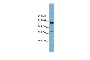
Figure 1. Western blot analysis of ABCB4 using anti-ABCB4 antibody (PB9275). Electrophoresis was performed on a 5-20% SDS-PAGE gel at 70V (Stacking gel) / 90V (Resolving gel) for 2-3 hours. The sample well of each lane was loaded with 30 ug of sample under reducing conditions. Lane 1: human HepG2 whole cell lysates, Lane 2: rat liver tissue lysates, Lane 3: rat RH35 whole cell lysates, Lane 4: mouse liver tissue lysates. After electrophoresis, proteins were transferred to a nitrocellulose membrane at 150 mA for 50-90 minutes. Blocked the membrane with 5% non-fat milk/TBS for 1.5 hour at RT. The membrane was incubated with rabbit anti-ABCB4 antigen affinity purified polyclonal antibody (Catalog # PB9275) at 0.5 microg/mL overnight at 4°C, then washed with TBS-0.1%Tween 3 times with 5 minutes each and probed with a goat anti-rabbit IgG-HRP secondary antibody at a dilution of 1:5000 for 1.5 hour at RT. The signal is developed using an Enhanced Chemiluminescent detection (ECL) kit (Catalog # EK1002) with Tanon 5200 system. A specific band was detected for ABCB4 at approximately 150 kDa. The expected band size for ABCB4 is at 142 kDa.
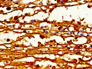
. ABCB4 was detected in a paraffin-embedded section of human liver cancer tissue. Heat mediated antigen retrieval was performed in EDTA buffer (pH 8.0, epitope retrieval solution). The tissue section was blocked with 10% goat serum. The tissue section was then incubated with 2 microg/ml rabbit anti-ABCB4 Antibody (PB9275) overnight at 4°C. Peroxidase Conjugated Goat Anti-rabbit IgG was used as secondary antibody and incubated for 30 minutes at 37°C. The tissue section was developed using HRP Conjugated Rabbit IgG Super Vision Assay Kit (Catalog # SV0002) with DAB as the chromogen.")
. Overlay histogram showing HepG2 cells stained with PB9275 (Blue line). To facilitate intracellular staining, cells were fixed with 4% paraformaldehyde and permeabilized with permeabilization buffer. The cells were blocked with 10% normal goat serum. And then incubated with rabbit anti-ABCB4 Antibody (PB9275, 1 microg/1x106 cells) for 30 min at 20°C. DyLight®488 conjugated goat anti-rabbit IgG (BA1127, 5-10 microg/1x106 cells) was used as secondary antibody for 30 minutes at 20°C. Isotype control antibody (Green line) was rabbit IgG (1 microg/1x106) used under the same conditions. Unlabelled sample without incubation with primary antibody and secondary antibody (Red line) was used as a blank control.")
Figure 1. Western blot analysis of ABCB4 using anti-ABCB4 antibody (PB9275). Electrophoresis was performed on a 5-20% SDS-PAGE gel at 70V (Stacking gel) / 90V (Resolving gel) for 2-3 hours. The sample well of each lane was loaded with 30 ug of sample under reducing conditions. Lane 1: human HepG2 whole cell lysates, Lane 2: rat liver tissue lysates, Lane 3: rat RH35 whole cell lysates, Lane 4: mouse liver tissue lysates. After electrophoresis, proteins were transferred to a nitrocellulose membrane at 150 mA for 50-90 minutes. Blocked the membrane with 5% non-fat milk/TBS for 1.5 hour at RT. The membrane was incubated with rabbit anti-ABCB4 antigen affinity purified polyclonal antibody (Catalog # PB9275) at 0.5 microg/mL overnight at 4°C, then washed with TBS-0.1%Tween 3 times with 5 minutes each and probed with a goat anti-rabbit IgG-HRP secondary antibody at a dilution of 1:5000 for 1.5 hour at RT. The signal is developed using an Enhanced Chemiluminescent detection (ECL) kit (Catalog # EK1002) with Tanon 5200 system. A specific band was detected for ABCB4 at approximately 150 kDa. The expected band size for ABCB4 is at 142 kDa.
Anti-ABCB4 Antibody Picoband(r)
PB9275
ApplicationsFlow Cytometry, Western Blot, ImmunoHistoChemistry
Product group Antibodies
ReactivityHuman, Mouse, Rat
TargetABCB4
Overview
- SupplierBoster Bio
- Product NameAnti-ABCB4 Antibody Picoband(r)
- Delivery Days Customer9
- Application Supplier NoteWB: The detection limit for ABCB4 is approximately 0.1ng/lane under reducing conditions. Tested Species: In-house tested species with positive results. By Heat: Boiling the paraffin sections in 10mM citrate buffer, pH6.0, for 20mins is required for the staining of formalin/paraffin sections. Other applications have not been tested. Optimal dilutions should be determined by end users.
- ApplicationsFlow Cytometry, Western Blot, ImmunoHistoChemistry
- CertificationResearch Use Only
- ClonalityPolyclonal
- Concentration500 ug/ml
- Gene ID5244
- Target nameABCB4
- Target descriptionATP binding cassette subfamily B member 4
- Target synonymsABC21, GBD1, ICP3, MDR2, MDR2/3, MDR3, PFIC-3, PGY3, phosphatidylcholine translocator ABCB4, ATP-binding cassette sub-family B member 4, ATP-binding cassette, sub-family B (MDR/TAP), member 4, P-glycoprotein 3, P-glycoprotein-3/multiple drug resistance-3, multidrug resistance protein 3, multiple drug resistance 3
- HostRabbit
- IsotypeIgG
- Protein IDP21439
- Protein NamePhosphatidylcholine translocator ABCB4
- Scientific DescriptionBoster Bio Anti-ABCB4 Antibody Picoband® catalog # PB9275. Tested in Flow Cytometry, IHC, WB applications. This antibody reacts with Human, Mouse, Rat. The brand Picoband indicates this is a premium antibody that guarantees superior quality, high affinity, and strong signals with minimal background in Western blot applications. Only our best-performing antibodies are designated as Picoband, ensuring unmatched performance.
- ReactivityHuman, Mouse, Rat
- Storage Instruction-20°C,2°C to 8°C
- UNSPSC12352203
Datasheet
MSDS
Related products
Product group Antibodies
ABCB4 AntibodyCSB-PA001050LA01HU
ApplicationsImmunoFluorescence, ELISA, ImmunoHistoChemistry
ReactivityHuman
TargetABCB4
- SizePrice
Product group Antibodies
Anti-ABCB4 AntibodyA326208
ApplicationsELISA, ImmunoHistoChemistry
ReactivityHuman
- SizePrice
Product group Antibodies

ABCB4 / MDR3 AntibodyLS-C832675
ApplicationsELISA, ImmunoHistoChemistry
ReactivityHuman
TargetABCB4
- SizePrice

Product group Antibodies
Anti-ABCB4 AntibodyHPA053288
ApplicationsImmunoHistoChemistry
ReactivityHuman
TargetABCB4
- SizePrice
Product group Antibodies

Goat anti-MDR2 / MDR3EB08703
ApplicationsELISA, ImmunoHistoChemistry
ReactivityHuman
TargetABCB4
- SizePrice
Product group Antibodies
ApplicationsImmunoPrecipitation, Western Blot, ImmunoCytoChemistry, ImmunoHistoChemistry
TargetABCB4
- SizePrice
Product group Antibodies
Anti-ABCB4 Antibody Picoband(r)PB9275-CARRIER-FREE
ApplicationsFlow Cytometry, Western Blot, ImmunoHistoChemistry
ReactivityHuman, Mouse, Rat
TargetABCB4
- SizePrice
Product group Antibodies
References
ABCB4 antibody, InternalGTX47122
ApplicationsWestern Blot
ReactivityHuman
TargetABCB4
- SizePrice
Product group Antibodies
Anti-ABCB4 Antibody144-66805
ApplicationsImmunoFluorescence, Western Blot
ReactivityHuman, Mouse
TargetABCB4
- SizePrice