


Immunofluorescent staining of human cell line Hep G2 shows localization to nucleus.
Immunofluorescent staining of human cell line Hep G2 shows localization to nucleus.
Anti-ABCG5 Antibody
HPA016514
ApplicationsImmunoCytoChemistry
Product group Antibodies
ReactivityHuman
TargetABCG5
Overview
- SupplierAtlas Antibodies
- Product NameAnti-ABCG5 Antibody
- Delivery Days Customer4
- ApplicationsImmunoCytoChemistry
- CertificationResearch Use Only
- ClonalityPolyclonal
- ConjugateUnconjugated
- Gene ID64240
- Target nameABCG5
- Target descriptionATP binding cassette subfamily G member 5
- Target synonymsSTSL, STSL2, ATP-binding cassette sub-family G member 5, ATP-binding cassette, sub-family G (WHITE), member 5, sterolin 1
- HostRabbit
- IsotypeIgG
- Protein IDQ9H222
- Protein NameATP-binding cassette sub-family G member 5
- Scientific DescriptionRecombinant Protein Epitope Signature Tag (PrEST) antigen sequence
- ReactivityHuman
- Storage Instruction-20°C,2°C to 8°C
- UNSPSC41116161
Datasheet
MSDS
Related products
Product group Antibodies

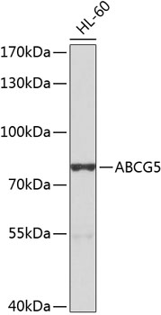
ABCG5 AntibodyLS-C410124
ApplicationsWestern Blot
ReactivityHuman
TargetABCG5
- SizePrice
Product group Antibodies
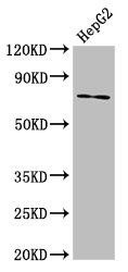
ABCG5 AntibodyCSB-PA863956LA01HU
ApplicationsImmunoFluorescence, Western Blot, ELISA, ImmunoHistoChemistry
ReactivityHuman
TargetABCG5
- SizePrice
Product group Antibodies
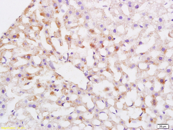
Abcg5 Polyclonal AntibodyCAC11286
ApplicationsImmunoFluorescence, Western Blot, ELISA, ImmunoHistoChemistry
TargetABCG5
- SizePrice

Product group Antibodies
Anti-ABCG5 Antibody Picoband(r)PB9415-CARRIER-FREE
ApplicationsWestern Blot
ReactivityChicken, Human
TargetABCG5
- SizePrice
Product group Antibodies
References
ABCG5 Polyclonal AntibodyBS-5013R
ApplicationsImmunoFluorescence, Western Blot, ELISA, ImmunoCytoChemistry, ImmunoHistoChemistry, ImmunoHistoChemistry Frozen, ImmunoHistoChemistry Paraffin
ReactivityHuman
TargetABCG5
- SizePrice
![FACS analysis of A549 cells using GTX60788 ABCG5 antibody [1B5E10]. Green : ABCG5 Red : negative control](https://www.genetex.com/upload/website/prouct_img/normal/GTX60788/GTX60788_20170912_FACS_w_23061123_410.webp)
Product group Antibodies
ABCG5 antibody [1B5E10]GTX60788
ApplicationsFlow Cytometry, Western Blot, ELISA, ImmunoHistoChemistry, ImmunoHistoChemistry Paraffin
ReactivityHuman
TargetABCG5
- SizePrice