


Immunohistochemistry analysis in human kidney and tonsil tissues using AMAb91259 antibody. Corresponding ACE2 RNA-seq data are presented for the same tissues.

Immunohistochemistry analysis in human kidney and tonsil tissues using AMAb91259 antibody. Corresponding ACE2 RNA-seq data are presented for the same tissues.
Anti-ACE2 Antibody
AMAB91259
ApplicationsWestern Blot, ImmunoHistoChemistry
Product group Antibodies
ReactivityHuman
TargetACE2
Overview
- SupplierAtlas Antibodies
- Product NameAnti-ACE2 Antibody
- Delivery Days Customer4
- ApplicationsWestern Blot, ImmunoHistoChemistry
- CertificationResearch Use Only
- ClonalityMonoclonal
- Clone IDCL4013
- ConjugateUnconjugated
- Gene ID59272
- Target nameACE2
- Target descriptionangiotensin converting enzyme 2
- Target synonymsACEH, angiotensin-converting enzyme 2, ACE-related carboxypeptidase, angiotensin I converting enzyme (peptidyl-dipeptidase A) 2, angiotensin I converting enzyme 2, angiotensin-converting enzyme homolog, angiotensin-converting enzyme-related carboxypeptidase, metalloprotease MPROT15, peptidyl-dipeptidase A
- HostMouse
- IsotypeIgG1
- Protein IDQ9BYF1
- Protein NameAngiotensin-converting enzyme 2
- Scientific DescriptionRecombinant Protein Epitope Signature Tag (PrEST) antigen sequence
- ReactivityHuman
- Storage Instruction-20°C,2°C to 8°C
- UNSPSC41116161
Datasheet
MSDS
Related products
Product group Antibodies
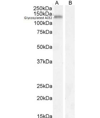
Anti-ACE2 AntibodyA286092
ApplicationsWestern Blot, ELISA, ImmunoHistoChemistry
ReactivityHuman
- SizePrice
Product group Antibodies
anti-ACE2 (human), pAbAG-25A-0042
ApplicationsWestern Blot, ELISA
ReactivityHuman
TargetACE2
- SizePrice

Product group Antibodies
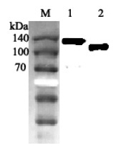
Anti-ACE2 Antibody Picoband(r)A00756-3-CARRIER-FREE
ApplicationsFlow Cytometry, Western Blot, ELISA
ReactivityHuman
TargetACE2
- SizePrice
Product group Antibodies
Anti-ACE2 Antibody130-10857
ApplicationsELISA
ReactivityVirus
TargetACE2
- SizePrice
Product group Antibodies

Anti-ACE2 [h11B11]AB03703-1.1
ApplicationsFlow Cytometry, ELISA, Neutralisation/Blocking
ReactivityHuman, Monkey
TargetACE2
- SizePrice

Product group Antibodies
Anti-ACE2 AntibodyAMAB91262
ApplicationsWestern Blot, ImmunoHistoChemistry
ReactivityHuman
TargetACE2
- SizePrice
Product group Antibodies
References
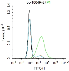
ACE2 Polyclonal AntibodyBS-1004R
ApplicationsFlow Cytometry, ImmunoFluorescence, Western Blot, ELISA, ImmunoCytoChemistry, ImmunoHistoChemistry, ImmunoHistoChemistry Frozen, ImmunoHistoChemistry Paraffin
ReactivityHuman
TargetACE2
- SizePrice
Product group Antibodies

Goat anti-ACE2 (C Terminal)EB13085
ApplicationsWestern Blot, ELISA, ImmunoHistoChemistry
ReactivityHuman
TargetACE2
- SizePrice