


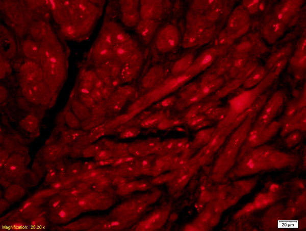
Immunofluorescent staining of human cell line MCF7 shows localization to the Golgi apparatus.
Immunofluorescent staining of human cell line MCF7 shows localization to the Golgi apparatus.
Anti-ACHE Antibody
HPA027098
ApplicationsImmunoCytoChemistry
Product group Antibodies
ReactivityHuman
TargetACHE
Overview
- SupplierAtlas Antibodies
- Product NameAnti-ACHE Antibody
- Delivery Days Customer4
- ApplicationsImmunoCytoChemistry
- CertificationResearch Use Only
- ClonalityPolyclonal
- ConjugateUnconjugated
- Gene ID43
- Target nameACHE
- Target descriptionacetylcholinesterase (Yt blood group)
- Target synonymsACEE, ARACHE, N-ACHE, YT, acetylcholinesterase, Yt blood group, acetylcholinesterase (Cartwright blood group), apoptosis-related acetylcholinesterase
- HostRabbit
- IsotypeIgG
- Protein IDP22303
- Protein NameAcetylcholinesterase
- Scientific DescriptionRecombinant Protein Epitope Signature Tag (PrEST) antigen sequence
- ReactivityHuman
- Storage Instruction-20°C,2°C to 8°C
- UNSPSC41116161
Datasheet
MSDS
Related products
Product group Antibodies
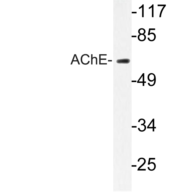
Anti-AChE AntibodyA96247
ApplicationsWestern Blot, ELISA, ImmunoHistoChemistry
ReactivityHuman, Mouse, Rat
- SizePrice
Product group Antibodies

Anti-acetylcholinesterase [1G]AB04029-1.1
ApplicationsELISA
ReactivityHuman
TargetACHE
- SizePrice
Product group Antibodies

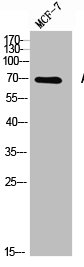
ACHE / Acetylcholinesterase AntibodyLS-C318075
ApplicationsWestern Blot, ELISA
ReactivityBovine
TargetACHE
- SizePrice
Product group Antibodies

Goat anti-ACHEEB06659
ApplicationsFlow Cytometry, ImmunoFluorescence, Western Blot, ELISA
ReactivityHuman, Mouse, Rat
TargetACHE
- SizePrice
Product group Antibodies
ACHE AntibodyCSB-PA010202
ApplicationsWestern Blot, ELISA, ImmunoHistoChemistry
ReactivityHuman, Mouse, Rat
TargetACHE
- SizePrice
Product group Antibodies
References
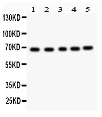
ACHE Polyclonal AntibodyBS-2511R
ApplicationsImmunoFluorescence, Western Blot, ELISA, ImmunoCytoChemistry, ImmunoHistoChemistry, ImmunoHistoChemistry Frozen, ImmunoHistoChemistry Paraffin
ReactivityBovine, Canine, Equine, Human, Mouse, Rat
TargetACHE
- SizePrice
Product group Antibodies
ApplicationsWestern Blot, ELISA, ImmunoCytoChemistry, ImmunoHistoChemistry, ImmunoHistoChemistry Frozen, ImmunoHistoChemistry Paraffin
ReactivityMouse, Rat
TargetACHE
- SizePrice
Product group Antibodies
Anti-Acetylcholinesterase/ACHE Antibody Picoband(r)PB9417-CARRIER-FREE
ApplicationsWestern Blot
ReactivityHamster, Human, Mouse, Rat
TargetACHE
- SizePrice