

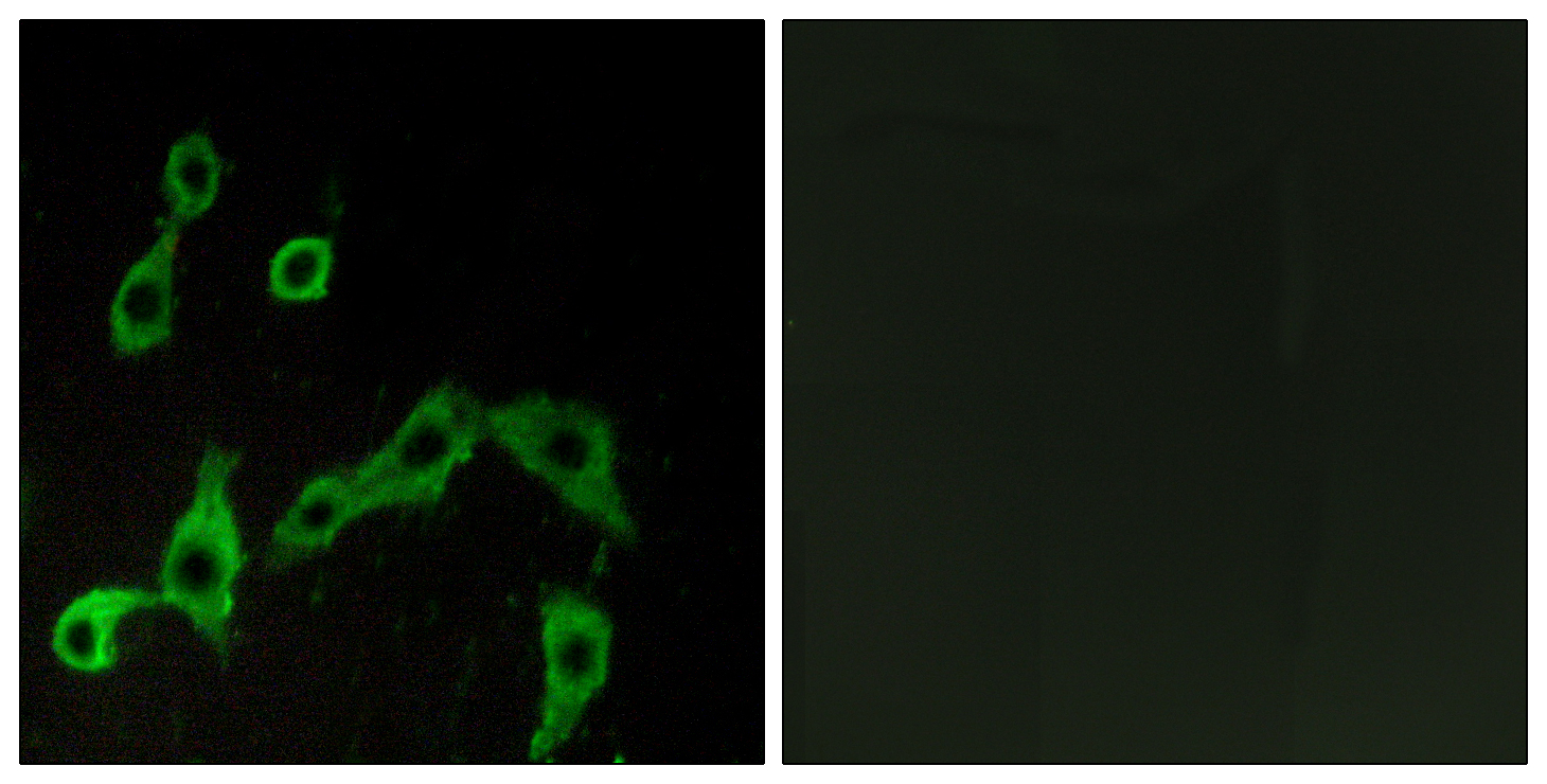

Anti-ADRA2A Antibody
A100622
ApplicationsImmunoFluorescence, ELISA
Product group Antibodies
ReactivityHuman
Overview
- SupplierAntibodies.com
- Product NameAnti-ADRA2A Antibody
- Delivery Days Customer7
- ApplicationsImmunoFluorescence, ELISA
- CertificationResearch Use Only
- ClonalityPolyclonal
- ConjugateUnconjugated
- HostRabbit
- IsotypeIgG
- Scientific DescriptionRabbit polyclonal antibody to ADRA2A.
- ReactivityHuman
- UNSPSC12352203
Related products
Product group Antibodies
ADRA2A AntibodyCSB-PA001388LA01HU
ApplicationsImmunoFluorescence, ELISA
ReactivityHuman
TargetADRA2A
- SizePrice

Product group Antibodies
Anti-alpha 2a Adrenergic Receptor/ADRA2A Antibody Picoband(r)A00883-3-CARRIER-FREE
ApplicationsFlow Cytometry, Western Blot, ELISA
ReactivityHuman, Mouse, Rat
TargetADRA2A
- SizePrice
Product group Antibodies

ADRA2A AntibodyLS-C748957
ApplicationsWestern Blot
ReactivityHuman, Mouse, Rat
TargetADRA2A
- SizePrice
Product group Antibodies

Goat anti-ADRA2AEB07121
ApplicationsFlow Cytometry, ImmunoFluorescence, ELISA
ReactivityCanine, Human, Mouse, Rat
TargetADRA2A
- SizePrice
![Boiled and unboiled LNCap whole cell and membrane extracts (30 μg) were separated by 10% SDS-PAGE, and the membrane was blotted with alpha 2a Adrenergic Receptor antibody [HL3723] (GTX641913) diluted at 1:1000. The HRP-conjugated anti-rabbit IgG antibody (GTX213110-01) was used to detect the primary antibody. (WCE: whole cell extract; ME: membrane extract)](https://www.genetex.com/upload/website/prouct_img/normal/GTX641913/GTX641913_T-45663_20250124_WB_Fraction_ub_25020422_270.webp)
Product group Antibodies
ApplicationsWestern Blot
ReactivityHuman, Mouse
TargetADRA2A
- SizePrice
Product group Antibodies
References
ADRA2 Polyclonal AntibodyBS-1062R
ApplicationsImmunoFluorescence, Western Blot, ELISA, ImmunoCytoChemistry, ImmunoHistoChemistry, ImmunoHistoChemistry Frozen, ImmunoHistoChemistry Paraffin
TargetADRA2A
- SizePrice
Product group Antibodies
Anti-ADRA2A Antibody144-02809
ApplicationsWestern Blot
ReactivityHuman, Mouse
TargetADRA2A
- SizePrice