


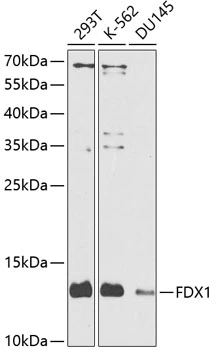
Figure 1. Western blot analysis of FDX1 using anti-FDX1 antibody (M05441). Electrophoresis was performed on a 5-20% SDS-PAGE gel at 70V (Stacking gel) / 90V (Resolving gel) for 2-3 hours. The sample well of each lane was loaded with 30 ug of sample under reducing conditions. Lane 1: human A549 whole cell lysates, Lane 2: human CACO-2 whole cell lysates, Lane 3: human SiHa whole cell lysates, Lane 4: human 293T whole cell lysates, Lane 5: rat PC-12 whole cell lysates, Lane 6: rat C6 whole cell lysates. After electrophoresis, proteins were transferred to a nitrocellulose membrane at 150 mA for 50-90 minutes. Blocked the membrane with 5% non-fat milk/TBS for 1.5 hour at RT. The membrane was incubated with rabbit anti-FDX1 antigen affinity purified monoclonal antibody (Catalog # M05441) at 1:500 overnight at 4°C, then washed with TBS-0.1%Tween 3 times with 5 minutes each and probed with a goat anti-rabbit IgG-HRP secondary antibody at a dilution of 1:1000 for 1.5 hour at RT. The signal is developed using an Enhanced Chemiluminescent detection (ECL) kit (Catalog # EK1002) with Tanon 5200 system. A specific band was detected for FDX1 at approximately 13 kDa. The expected band size for FDX1 is at 19 kDa.

Figure 1. Western blot analysis of FDX1 using anti-FDX1 antibody (M05441). Electrophoresis was performed on a 5-20% SDS-PAGE gel at 70V (Stacking gel) / 90V (Resolving gel) for 2-3 hours. The sample well of each lane was loaded with 30 ug of sample under reducing conditions. Lane 1: human A549 whole cell lysates, Lane 2: human CACO-2 whole cell lysates, Lane 3: human SiHa whole cell lysates, Lane 4: human 293T whole cell lysates, Lane 5: rat PC-12 whole cell lysates, Lane 6: rat C6 whole cell lysates. After electrophoresis, proteins were transferred to a nitrocellulose membrane at 150 mA for 50-90 minutes. Blocked the membrane with 5% non-fat milk/TBS for 1.5 hour at RT. The membrane was incubated with rabbit anti-FDX1 antigen affinity purified monoclonal antibody (Catalog # M05441) at 1:500 overnight at 4°C, then washed with TBS-0.1%Tween 3 times with 5 minutes each and probed with a goat anti-rabbit IgG-HRP secondary antibody at a dilution of 1:1000 for 1.5 hour at RT. The signal is developed using an Enhanced Chemiluminescent detection (ECL) kit (Catalog # EK1002) with Tanon 5200 system. A specific band was detected for FDX1 at approximately 13 kDa. The expected band size for FDX1 is at 19 kDa.
Anti-ADX Rabbit Monoclonal Antibody
M05441
ApplicationsImmunoFluorescence, Western Blot, ImmunoCytoChemistry, ImmunoHistoChemistry
Product group Antibodies
ReactivityHuman, Mouse, Rat
TargetFDX1
Overview
- SupplierBoster Bio
- Product NameAnti-ADX Rabbit Monoclonal Antibody
- Delivery Days Customer9
- ApplicationsImmunoFluorescence, Western Blot, ImmunoCytoChemistry, ImmunoHistoChemistry
- CertificationResearch Use Only
- ClonalityMonoclonal
- Clone ID28F80
- Gene ID2230
- Target nameFDX1
- Target descriptionferredoxin 1
- Target synonymsADX, FDX, LOH11CR1D, adrenodoxin, mitochondrial, adrenal ferredoxin, hepatoredoxin, mitochondrial adrenodoxin
- HostRabbit
- IsotypeIgG
- Protein IDP10109
- Protein NameAdrenodoxin, mitochondrial
- Scientific DescriptionBoster Bio Anti-ADX Rabbit Monoclonal Antibody catalog # M05441. Tested in WB, IHC, ICC/IF applications. This antibody reacts with Human, Mouse, Rat.
- ReactivityHuman, Mouse, Rat
- Storage Instruction-20°C
- UNSPSC12352203
References
- Li X, Wang J, Guo Z, et al. Copper metabolism-related risk score identifies hepatocellular carcinoma subtypes and SLC27A5 as a potential regulator of cuproptosis. Aging (Albany NY). 2023,15(24):15084-15113. doi: 10.18632/aging.205334Read this paper
- Yao Y, Chen H, Lou M, et al. Cuproptosis-related gene FDX1 as a prognostic biomarker for kidney renal clear cell carcinoma correlates with immune checkpoints and immune cell infiltration. Front Genet. 2023,14:1071694. doi: 10.3389/fgene.2023.1071694Read this paper
Related products
Product group Antibodies
FDX1 AntibodyCSB-PA008570ZA01BO
ApplicationsWestern Blot, ELISA
ReactivityBovine
TargetFDX1
- SizePrice

Product group Antibodies
Anti-FDX1 AntibodyHPA062087
ApplicationsImmunoCytoChemistry, ImmunoHistoChemistry
ReactivityHuman
TargetFDX1
- SizePrice
Product group Antibodies

FDX1 / ADX AntibodyLS-C411342
ApplicationsWestern Blot, ImmunoHistoChemistry
ReactivityHuman, Mouse, Rat
TargetFDX1
- SizePrice
Product group Antibodies
Adrenodoxin antibodyGTX65989
ApplicationsWestern Blot
ReactivityHuman
TargetFDX1
- SizePrice
Product group Antibodies
Adrenodoxin Polyclonal AntibodyBS-11426R
ApplicationsImmunoFluorescence, Western Blot, ELISA, ImmunoCytoChemistry, ImmunoHistoChemistry, ImmunoHistoChemistry Frozen, ImmunoHistoChemistry Paraffin
ReactivityHuman, Mouse, Rat, Sheep
TargetFDX1
- SizePrice
Product group Antibodies
Anti-FDX1 Antibody144-09815
ApplicationsWestern Blot, ImmunoHistoChemistry
ReactivityHuman, Mouse, Rat
TargetFDX1
- SizePrice