


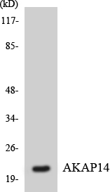
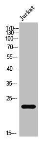
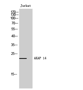
Figure 1. Western blot analysis of AKAP14 using anti-AKAP14 antibody (M15886). Electrophoresis was performed on a 5-20% SDS-PAGE gel at 70V (Stacking gel) / 90V (Resolving gel) for 2-3 hours. The sample well of each lane was loaded with 30 ug of sample under reducing conditions. Lane 1: human Jurkat whole cell lysates, Lane 2: human A549 whole cell lysates, Lane 3: human HepG2 whole cell lysates, Lane 4: human RT4 whole cell lysates, Lane 5: rat testis tissue lysates, Lane 6: rat C6 whole cell lysates, Lane 7: mouse testis tissue lysates, Lane 8: mouse Neuro-2a whole cell lysates. After electrophoresis, proteins were transferred to a nitrocellulose membrane at 150 mA for 50-90 minutes. Blocked the membrane with 5% non-fat milk/TBS for 1.5 hour at RT. The membrane was incubated with rabbit anti-AKAP14 antigen affinity purified monoclonal antibody (Catalog # M15886) at 1:500 overnight at 4°C, then washed with TBS-0.1%Tween 3 times with 5 minutes each and probed with a goat anti-rabbit IgG-HRP secondary antibody at a dilution of 1:500 for 1.5 hour at RT. The signal is developed using an Enhanced Chemiluminescent detection (ECL) kit (Catalog # EK1002) with Tanon 5200 system. A specific band was detected for AKAP14 at approximately 21,23 kDa. The expected band size for AKAP14 is at 21 kDa.
Figure 1. Western blot analysis of AKAP14 using anti-AKAP14 antibody (M15886). Electrophoresis was performed on a 5-20% SDS-PAGE gel at 70V (Stacking gel) / 90V (Resolving gel) for 2-3 hours. The sample well of each lane was loaded with 30 ug of sample under reducing conditions. Lane 1: human Jurkat whole cell lysates, Lane 2: human A549 whole cell lysates, Lane 3: human HepG2 whole cell lysates, Lane 4: human RT4 whole cell lysates, Lane 5: rat testis tissue lysates, Lane 6: rat C6 whole cell lysates, Lane 7: mouse testis tissue lysates, Lane 8: mouse Neuro-2a whole cell lysates. After electrophoresis, proteins were transferred to a nitrocellulose membrane at 150 mA for 50-90 minutes. Blocked the membrane with 5% non-fat milk/TBS for 1.5 hour at RT. The membrane was incubated with rabbit anti-AKAP14 antigen affinity purified monoclonal antibody (Catalog # M15886) at 1:500 overnight at 4°C, then washed with TBS-0.1%Tween 3 times with 5 minutes each and probed with a goat anti-rabbit IgG-HRP secondary antibody at a dilution of 1:500 for 1.5 hour at RT. The signal is developed using an Enhanced Chemiluminescent detection (ECL) kit (Catalog # EK1002) with Tanon 5200 system. A specific band was detected for AKAP14 at approximately 21,23 kDa. The expected band size for AKAP14 is at 21 kDa.
Anti-AKAP14 Rabbit Monoclonal Antibody
M15886
ApplicationsImmunoFluorescence, ImmunoPrecipitation, Western Blot, ImmunoCytoChemistry
Product group Antibodies
ReactivityHuman, Mouse, Rat
TargetAKAP14
Overview
- SupplierBoster Bio
- Product NameAnti-AKAP14 Rabbit Monoclonal Antibody
- Delivery Days Customer9
- ApplicationsImmunoFluorescence, ImmunoPrecipitation, Western Blot, ImmunoCytoChemistry
- CertificationResearch Use Only
- ClonalityMonoclonal
- Clone IDFGO-1
- Gene ID158798
- Target nameAKAP14
- Target descriptionA-kinase anchoring protein 14
- Target synonymsAKAP28, PRKA14, A-kinase anchor protein 14, A kinase (PRKA) anchor protein 14, A-kinase anchor protein 28 kDa, A-kinase anchoring protein 28, AKAP 28, AKAP-14, protein kinase A-anchoring protein 14
- HostRabbit
- IsotypeIgG
- Protein IDQ86UN6
- Protein NameA-kinase anchor protein 14
- Scientific DescriptionBoster Bio Anti-AKAP14 Rabbit Monoclonal Antibody catalog # M15886. Tested in WB, ICC/IF, IP applications. This antibody reacts with Human, Mouse, Rat.
- ReactivityHuman, Mouse, Rat
- Storage Instruction-20°C
- UNSPSC12352203
Datasheet
MSDS
Related products
Product group Antibodies
Anti-AKAP14 AntibodyA100618
ApplicationsWestern Blot, ELISA
ReactivityHuman
- SizePrice
Product group Antibodies
AKAP14 Recombinant Antibody, Biotin ConjugatedBSM-61280R-BIOTIN
ApplicationsImmunoPrecipitation, Western Blot
TargetAKAP14
- SizePrice
Product group Antibodies
AKAP14 AntibodyCSB-PA007591
ApplicationsImmunoFluorescence, Western Blot, ELISA, ImmunoHistoChemistry
ReactivityHuman
TargetAKAP14
- SizePrice
Product group Antibodies

AKAP14 Antibody (10-90 aa, N-terminal)LS-C381949
ApplicationsImmunoFluorescence, Western Blot, ELISA, ImmunoHistoChemistry
ReactivityHuman
TargetAKAP14
- SizePrice

Product group Antibodies
Anti-AKAP14 AntibodyHPA060999
ApplicationsImmunoHistoChemistry
ReactivityHuman
TargetAKAP14
- SizePrice
Product group Antibodies
AKAP14 antibodyGTX34363
ApplicationsWestern Blot
ReactivityHuman
TargetAKAP14
- SizePrice