


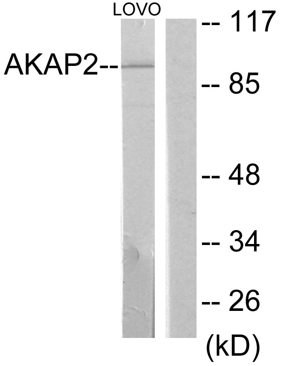
Figure 1. Western blot analysis of AKAP2 using anti-AKAP2 antibody (A09279). Electrophoresis was performed on a 5-20% SDS-PAGE gel at 70V (Stacking gel) / 90V (Resolving gel) for 2-3 hours. The sample well of each lane was loaded with 30 ug of sample under reducing conditions. Lane 1: human PC-3 whole cell lysates, Lane 2: mouse lung tissue lysates. After electrophoresis, proteins were transferred to a nitrocellulose membrane at 150 mA for 50-90 minutes. Blocked the membrane with 5% non-fat milk/TBS for 1.5 hour at RT. The membrane was incubated with rabbit anti-AKAP2 antigen affinity purified polyclonal antibody (Catalog # A09279) at 0.5 microg/mL overnight at 4°C, then washed with TBS-0.1%Tween 3 times with 5 minutes each and probed with a goat anti-rabbit IgG-HRP secondary antibody at a dilution of 1:5000 for 1.5 hour at RT. The signal is developed using an Enhanced Chemiluminescent detection (ECL) kit (Catalog # EK1002) with Tanon 5200 system. A specific band was detected for AKAP2 at approximately 150-170 kDa. The expected band size for AKAP2 is at 95 kDa.
Figure 1. Western blot analysis of AKAP2 using anti-AKAP2 antibody (A09279). Electrophoresis was performed on a 5-20% SDS-PAGE gel at 70V (Stacking gel) / 90V (Resolving gel) for 2-3 hours. The sample well of each lane was loaded with 30 ug of sample under reducing conditions. Lane 1: human PC-3 whole cell lysates, Lane 2: mouse lung tissue lysates. After electrophoresis, proteins were transferred to a nitrocellulose membrane at 150 mA for 50-90 minutes. Blocked the membrane with 5% non-fat milk/TBS for 1.5 hour at RT. The membrane was incubated with rabbit anti-AKAP2 antigen affinity purified polyclonal antibody (Catalog # A09279) at 0.5 microg/mL overnight at 4°C, then washed with TBS-0.1%Tween 3 times with 5 minutes each and probed with a goat anti-rabbit IgG-HRP secondary antibody at a dilution of 1:5000 for 1.5 hour at RT. The signal is developed using an Enhanced Chemiluminescent detection (ECL) kit (Catalog # EK1002) with Tanon 5200 system. A specific band was detected for AKAP2 at approximately 150-170 kDa. The expected band size for AKAP2 is at 95 kDa.
Anti-AKAP2 Antibody Picoband(r)
A09279-CARRIER-FREE
ApplicationsWestern Blot
Product group Antibodies
ReactivityHuman, Mouse
TargetAKAP2
Overview
- SupplierBoster Bio
- Product NameAnti-AKAP2 Antibody Picoband(r)
- Delivery Days Customer9
- Application Supplier NoteTested Species: In-house tested species with positive results. Other applications have not been tested. Optimal dilutions should be determined by end users.
- ApplicationsWestern Blot
- CertificationResearch Use Only
- ClonalityPolyclonal
- Concentration500 ug/ml
- Gene ID11217
- Target nameAKAP2
- Target descriptionA-kinase anchoring protein 2
- Target synonymsAKAP-2, AKAPKL, MISP2, PRKA2, A kinase (PRKA) anchor protein 2, protein kinase A anchoring protein 2, protein kinase A2
- HostRabbit
- IsotypeIgG
- Protein IDQ9Y2D5
- Protein NamePALM2-AKAP2 fusion protein
- Scientific DescriptionBoster Bio Anti-AKAP2 Antibody Picoband® catalog # A09279. Tested in WB applications. This antibody reacts with Human, Mouse. The brand Picoband indicates this is a premium antibody that guarantees superior quality, high affinity, and strong signals with minimal background in Western blot applications. Only our best-performing antibodies are designated as Picoband, ensuring unmatched performance.
- ReactivityHuman, Mouse
- Storage Instruction-20°C,2°C to 8°C
- UNSPSC12352203
Related products

Product group Antibodies
AKAP2/PALM2 Polyclonal AntibodyBS-9028R
ApplicationsImmunoFluorescence, Western Blot, ELISA, ImmunoHistoChemistry, ImmunoHistoChemistry Frozen, ImmunoHistoChemistry Paraffin
ReactivityHuman, Mouse, Rat
TargetAKAP2
- SizePrice
Product group Antibodies
AKAP2 AntibodyCSB-PA010254
ApplicationsWestern Blot, ELISA
ReactivityHuman
TargetAKAP2
- SizePrice
Product group Antibodies
Anti-AKAP2 AntibodyA101226
ApplicationsWestern Blot, ELISA
ReactivityHuman
- SizePrice

Product group Antibodies
AKAP2 antibodyGTX87891
ApplicationsWestern Blot
ReactivityHuman
TargetAKAP2
- SizePrice
Product group Antibodies
AKAP2 AntibodyPACO04361
ApplicationsWestern Blot, ELISA
ReactivityHuman
TargetAKAP2
- SizePrice
Product group Antibodies

AKAP2 Antibody (aa525-575)LS-C286058
ApplicationsImmunoPrecipitation
ReactivityHuman
TargetAKAP2
- SizePrice