


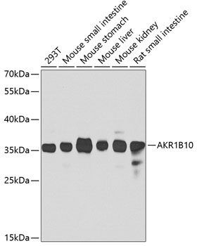
Figure 1. Western blot analysis of AKR1B10 using anti-AKR1B10 antibody (A02976). Electrophoresis was performed on a 5-20% SDS-PAGE gel at 70V (Stacking gel) / 90V (Resolving gel) for 2-3 hours. The sample well of each lane was loaded with 30 ug of sample under reducing conditions. Lane 1: human A549 whole cell lysates, Lane 2: human HepG2 whole cell lysates. After electrophoresis, proteins were transferred to a nitrocellulose membrane at 150 mA for 50-90 minutes. Blocked the membrane with 5% non-fat milk/TBS for 1.5 hour at RT. The membrane was incubated with rabbit anti-AKR1B10 antigen affinity purified polyclonal antibody (Catalog # A02976) at 0.5 microg/mL overnight at 4°C, then washed with TBS-0.1%Tween 3 times with 5 minutes each and probed with a goat anti-rabbit IgG-HRP secondary antibody at a dilution of 1:5000 for 1.5 hour at RT. The signal is developed using an Enhanced Chemiluminescent detection (ECL) kit (Catalog # EK1002) with Tanon 5200 system. A specific band was detected for AKR1B10 at approximately 36 kDa. The expected band size for AKR1B10 is at 36 kDa.
. AKR1B10 was detected in paraffin-embedded section of human intestinal cancer tissues. Heat mediated antigen retrieval was performed in citrate buffer (pH6, epitope retrieval solution) for 20 mins. The tissue section was blocked with 10% goat serum. The tissue section was then incubated with 1microg/ml rabbit anti-AKR1B10 Antibody (A02976) overnight at 4°C. Biotinylated goat anti-rabbit IgG was used as secondary antibody and incubated for 30 minutes at 37°C. The tissue section was developed using Strepavidin-Biotin-Complex (SABC)(Catalog # SA1022) with DAB as the chromogen.")
. AKR1B10 was detected in paraffin-embedded section of human liver cancer tissues. Heat mediated antigen retrieval was performed in citrate buffer (pH6, epitope retrieval solution) for 20 mins. The tissue section was blocked with 10% goat serum. The tissue section was then incubated with 1microg/ml rabbit anti-AKR1B10 Antibody (A02976) overnight at 4°C. Biotinylated goat anti-rabbit IgG was used as secondary antibody and incubated for 30 minutes at 37°C. The tissue section was developed using Strepavidin-Biotin-Complex (SABC)(Catalog # SA1022) with DAB as the chromogen.")
. AKR1B10 was detected in an immunocytochemical section of A549 cells. Enzyme antigen retrieval was performed using IHC enzyme antigen retrieval reagent (AR0022) for 15 mins. The cells were blocked with 10% goat serum. And then incubated with 5 microg/mL rabbit anti-AKR1B10 Antibody (A02976) overnight at 4°C. DyLight®488 Conjugated Goat Anti-Rabbit IgG (BA1127) was used as secondary antibody at 1:500 dilution and incubated for 30 minutes at 37°C. The section was counterstained with DAPI. Visualize using a fluorescence microscope and filter sets appropriate for the label used.")
Figure 1. Western blot analysis of AKR1B10 using anti-AKR1B10 antibody (A02976). Electrophoresis was performed on a 5-20% SDS-PAGE gel at 70V (Stacking gel) / 90V (Resolving gel) for 2-3 hours. The sample well of each lane was loaded with 30 ug of sample under reducing conditions. Lane 1: human A549 whole cell lysates, Lane 2: human HepG2 whole cell lysates. After electrophoresis, proteins were transferred to a nitrocellulose membrane at 150 mA for 50-90 minutes. Blocked the membrane with 5% non-fat milk/TBS for 1.5 hour at RT. The membrane was incubated with rabbit anti-AKR1B10 antigen affinity purified polyclonal antibody (Catalog # A02976) at 0.5 microg/mL overnight at 4°C, then washed with TBS-0.1%Tween 3 times with 5 minutes each and probed with a goat anti-rabbit IgG-HRP secondary antibody at a dilution of 1:5000 for 1.5 hour at RT. The signal is developed using an Enhanced Chemiluminescent detection (ECL) kit (Catalog # EK1002) with Tanon 5200 system. A specific band was detected for AKR1B10 at approximately 36 kDa. The expected band size for AKR1B10 is at 36 kDa.
Anti-AKR1B10 Antibody Picoband(r)
A02976-CARRIER-FREE
ApplicationsImmunoFluorescence, Western Blot, ImmunoCytoChemistry, ImmunoHistoChemistry
Product group Antibodies
ReactivityHuman
TargetAKR1B10
Overview
- SupplierBoster Bio
- Product NameAnti-AKR1B10 Antibody Picoband(r)
- Delivery Days Customer9
- Application Supplier NoteTested Species: In-house tested species with positive results. By Heat: Boiling the paraffin sections in 10mM citrate buffer, pH6.0, for 20mins is required for the staining of formalin/paraffin sections. Other applications have not been tested. Optimal dilutions should be determined by end users.
- ApplicationsImmunoFluorescence, Western Blot, ImmunoCytoChemistry, ImmunoHistoChemistry
- CertificationResearch Use Only
- ClonalityPolyclonal
- Concentration500 ug/ml
- Gene ID57016
- Target nameAKR1B10
- Target descriptionaldo-keto reductase family 1 member B10
- Target synonymsAKR1B11, AKR1B12, ALDRLn, ARL-1, ARL1, HIS, HSI, aldo-keto reductase family 1 member B10, ARP, SI reductase, aldo-keto reductase family 1, member B10 (aldose reductase), aldo-keto reductase family 1, member B11 (aldose reductase-like), aldose reductase-like 1, aldose reductase-like peptide, aldose reductase-related protein, hARP, small intestine reductase
- HostRabbit
- IsotypeIgG
- Protein IDO60218
- Protein NameAldo-keto reductase family 1 member B10
- Scientific DescriptionBoster Bio Anti-AKR1B10 Antibody Picoband® catalog # A02976. Tested in IF, IHC, ICC, WB applications. This antibody reacts with Human. The brand Picoband indicates this is a premium antibody that guarantees superior quality, high affinity, and strong signals with minimal background in Western blot applications. Only our best-performing antibodies are designated as Picoband, ensuring unmatched performance.
- ReactivityHuman
- Storage Instruction-20°C,2°C to 8°C
- UNSPSC12352203
Related products
Product group Antibodies
Anti-AKR1B10 AntibodyA15917
ApplicationsWestern Blot
ReactivityHuman, Mouse, Rat
- SizePrice
Product group Antibodies
Anti-AKR1B10 Antibody144-07823
ApplicationsWestern Blot
ReactivityHuman, Mouse, Rat
TargetAKR1B10
- SizePrice
Product group Antibodies
AKR1B10 AntibodyCSB-PA001540ESR2HU
ApplicationsWestern Blot, ELISA, ImmunoHistoChemistry
ReactivityHuman
TargetAKR1B10
- SizePrice
Product group Antibodies

Goat anti-AKR1B10EB08336
ApplicationsWestern Blot, ELISA
ReactivityHuman
TargetAKR1B10
- SizePrice
Product group Antibodies
Akr1B10 Polyclonal AntibodyCAC10647
ApplicationsWestern Blot, ELISA, ImmunoHistoChemistry
TargetAKR1B10
- SizePrice
Product group Antibodies

AKR1B10 AntibodyLS-C409372
ApplicationsWestern Blot, ImmunoHistoChemistry
ReactivityHuman, Mouse, Rat
TargetAKR1B10
- SizePrice

Product group Antibodies
Anti-AKR1B10 AntibodyHPA020280
ApplicationsWestern Blot, ImmunoCytoChemistry, ImmunoHistoChemistry
ReactivityHuman
TargetAKR1B10
- SizePrice
Product group Antibodies
AKR1B10 antibodyGTX109571
ApplicationsImmunoFluorescence, Western Blot, ImmunoCytoChemistry, ImmunoHistoChemistry, ImmunoHistoChemistry Paraffin
ReactivityHuman, Mouse, Rat
TargetAKR1B10
- SizePrice