


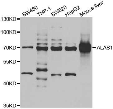
Figure 1. Western blot analysis of Alas1 using anti-Alas1 antibody (A10405-1). Electrophoresis was performed on a 5-20% SDS-PAGE gel at 70V (Stacking gel) / 90V (Resolving gel) for 2-3 hours. The sample well of each lane was loaded with 50ug of sample under reducing conditions. Lane 1: human U2OS whole cell lysates, Lane 2: human Hela whole cell lysates, Lane 3: human HepG2 whole cell lysates, Lane 4: rat NRK whole cell lysates, Lane 5: rat lung tissue lysates, Lane 6: mouse stomach tissue lysates, Lane 7: mouse NIH/3T3 whole cell lysates. After Electrophoresis, proteins were transferred to a Nitrocellulose membrane at 150mA for 50-90 minutes. Blocked the membrane with 5% Non-fat Milk/ TBS for 1.5 hour at RT. The membrane was incubated with rabbit anti-Alas1 antigen affinity purified polyclonal antibody (Catalog # A10405-1) at 0.5 microg/mL overnight at 4°C, then washed with TBS-0.1%Tween 3 times with 5 minutes each and probed with a goat anti-rabbit IgG-HRP secondary antibody at a dilution of 1:10000 for 1.5 hour at RT. The signal is developed using an Enhanced Chemiluminescent detection (ECL) kit (Catalog # EK1002) with Tanon 5200 system. A specific band was detected for Alas1 at approximately 71 kDa. The expected band size for Alas1 is at 71 kDa.
. Overlay histogram showing HEL cells stained with A10405-1 (Blue line). To facilitate intracellular staining, cells were fixed with 4% paraformaldehyde and permeabilized with permeabilization buffer. The cells were blocked with 10% normal goat serum. And then incubated with rabbit anti-Alas1 Antibody (A10405-1,1microg/1x106 cells) for 30 min at 20°C. DyLight®488 conjugated goat anti-rabbit IgG (BA1127, 5-10microg/1x106 cells) was used as secondary antibody for 30 minutes at 20°C. Isotype control antibody (Green line) was rabbit IgG (1microg/1x106) used under the same conditions. Unlabelled sample without incubation with primary antibody and secondary antibody (Red line) was used as a blank control.")
Figure 1. Western blot analysis of Alas1 using anti-Alas1 antibody (A10405-1). Electrophoresis was performed on a 5-20% SDS-PAGE gel at 70V (Stacking gel) / 90V (Resolving gel) for 2-3 hours. The sample well of each lane was loaded with 50ug of sample under reducing conditions. Lane 1: human U2OS whole cell lysates, Lane 2: human Hela whole cell lysates, Lane 3: human HepG2 whole cell lysates, Lane 4: rat NRK whole cell lysates, Lane 5: rat lung tissue lysates, Lane 6: mouse stomach tissue lysates, Lane 7: mouse NIH/3T3 whole cell lysates. After Electrophoresis, proteins were transferred to a Nitrocellulose membrane at 150mA for 50-90 minutes. Blocked the membrane with 5% Non-fat Milk/ TBS for 1.5 hour at RT. The membrane was incubated with rabbit anti-Alas1 antigen affinity purified polyclonal antibody (Catalog # A10405-1) at 0.5 microg/mL overnight at 4°C, then washed with TBS-0.1%Tween 3 times with 5 minutes each and probed with a goat anti-rabbit IgG-HRP secondary antibody at a dilution of 1:10000 for 1.5 hour at RT. The signal is developed using an Enhanced Chemiluminescent detection (ECL) kit (Catalog # EK1002) with Tanon 5200 system. A specific band was detected for Alas1 at approximately 71 kDa. The expected band size for Alas1 is at 71 kDa.
Anti-Alas1 Antibody Picoband(r)
A10405-1-DYLIGHT594
ApplicationsFlow Cytometry, Western Blot, ELISA
Product group Antibodies
ReactivityHuman, Mouse, Rat
TargetALAS1
Overview
- SupplierBoster Bio
- Product NameAnti-Alas1 Antibody Picoband(r)
- Delivery Days Customer9
- ApplicationsFlow Cytometry, Western Blot, ELISA
- CertificationResearch Use Only
- ClonalityPolyclonal
- Concentration500 ug/ml
- ConjugateOther Conjugate
- Gene ID211
- Target nameALAS1
- Target description5'-aminolevulinate synthase 1
- Target synonymsALAS, ALAS-H, ALAS3, ALASH, MIG4, 5-aminolevulinate synthase, non-specific, mitochondrial, 5-aminolevulinate synthase, nonspecific, mitochondrial, 5-aminolevulinic acid synthase 1, aminolevulinate, delta-, synthase 1, delta-ALA synthase 1, delta-aminolevulinate synthase 1, migration-inducing protein 4
- HostRabbit
- IsotypeIgG
- Protein IDP13196
- Protein Name5-aminolevulinate synthase, non-specific, mitochondrial
- Scientific DescriptionBoster Bio Anti-Alas1 Antibody Picoband® catalog # A10405-1. Tested in ELISA, Flow Cytometry, WB applications. This antibody reacts with Human, Mouse, Rat. The brand Picoband indicates this is a premium antibody that guarantees superior quality, high affinity, and strong signals with minimal background in Western blot applications. Only our best-performing antibodies are designated as Picoband, ensuring unmatched performance.
- ReactivityHuman, Mouse, Rat
- Storage Instruction-20°C,2°C to 8°C
- UNSPSC12352203
Related products

Product group Antibodies
Alas1 (1G11) Recombinant AntibodyBSM-52012R
ApplicationsFlow Cytometry, ImmunoFluorescence, Western Blot, ImmunoCytoChemistry, ImmunoHistoChemistry, ImmunoHistoChemistry Frozen, ImmunoHistoChemistry Paraffin
ReactivityHuman, Mouse
TargetALAS1
- SizePrice
Product group Antibodies
Alas1 Recombinant AntibodyCAC11977
ApplicationsWestern Blot, ELISA
TargetALAS1
- SizePrice
Product group Antibodies
Anti-ALAS1 AntibodyA29558
ApplicationsWestern Blot, ImmunoHistoChemistry
ReactivityHuman
- SizePrice
Product group Antibodies
Anti-ALAS1 Antibody144-06521
ApplicationsWestern Blot
ReactivityHuman, Mouse, Rat
TargetALAS1
- SizePrice
Product group Antibodies
ALAS-H antibody [N1N3]GTX114245
ApplicationsWestern Blot
ReactivityHuman
TargetALAS1
- SizePrice
Product group Antibodies

ALAS1 AntibodyLS-C748510
ApplicationsImmunoFluorescence, Western Blot
ReactivityHuman, Mouse, Rat
TargetALAS1
- SizePrice
Product group Antibodies
ALAS1 AntibodyCSB-PA001559LA01HU
ApplicationsELISA, ImmunoHistoChemistry
ReactivityHuman
TargetALAS1
- SizePrice