


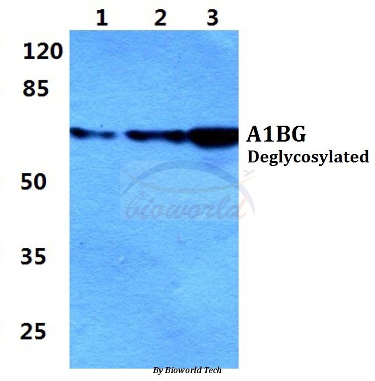
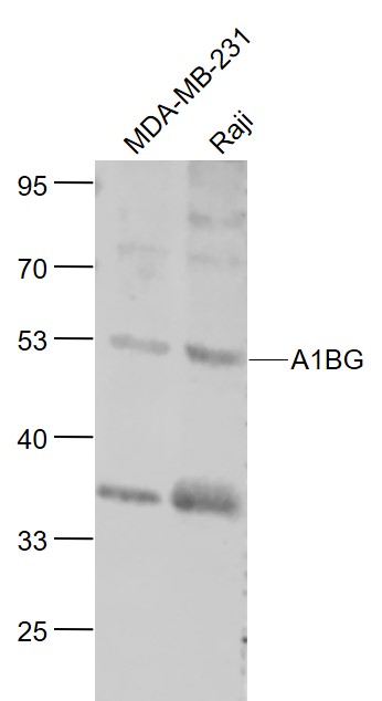
Figure 1. Western blot analysis of Alpha 1B-Glycoprotein/A1BG using anti-Alpha 1B-Glycoprotein/A1BG antibody (A07289-2). Electrophoresis was performed on a 5-20% SDS-PAGE gel at 70V (Stacking gel) / 90V (Resolving gel) for 2-3 hours. The sample well of each lane was loaded with 30 ug of sample under reducing conditions. Lane 1: human hepatocellular carcinoma tumor tissue (HCCT) lysates, Lane 2: human hepatocellular carcinoma paracancerous tissue (HCCP) lysates, Lane 3: rat plasma lysates, Lane 4: mouse plasma lysates. After electrophoresis, proteins were transferred to a nitrocellulose membrane at 150 mA for 50-90 minutes. Blocked the membrane with 5% non-fat milk/TBS for 1.5 hour at RT. The membrane was incubated with rabbit anti-Alpha 1B-Glycoprotein/A1BG antigen affinity purified polyclonal antibody (Catalog # A07289-2) at 0.25 microg/mL overnight at 4°C, then washed with TBS-0.1%Tween 3 times with 5 minutes each and probed with a goat anti-rabbit IgG-HRP secondary antibody at a dilution of 1:5000 for 1.5 hour at RT. The signal is developed using an Enhanced Chemiluminescent detection (ECL) kit (Catalog # EK1002) with Tanon 5200 system. A specific band was detected for Alpha 1B-Glycoprotein/A1BG at approximately 50-75 kDa. The expected band size for Alpha 1B-Glycoprotein/A1BG is at 54 kDa.
. Overlay histogram showing HepG2 cells stained with A07289-2 (Blue line). The cells were fixed with 4% paraformaldehyde and blocked with 10% normal goat serum. And then incubated with rabbit anti-Alpha 1B-Glycoprotein/A1BG Antibody (A07289-2, 1 microg/1x106 cells) for 30 min at 20°C. DyLight®488 conjugated goat anti-rabbit IgG (BA1127, 5-10 microg/1x106 cells) was used as secondary antibody for 30 minutes at 20°C. Isotype control antibody (Green line) was rabbit IgG (1 microg/1x106) used under the same conditions. Unlabelled sample (Red line) was also used as a control.")
Figure 1. Western blot analysis of Alpha 1B-Glycoprotein/A1BG using anti-Alpha 1B-Glycoprotein/A1BG antibody (A07289-2). Electrophoresis was performed on a 5-20% SDS-PAGE gel at 70V (Stacking gel) / 90V (Resolving gel) for 2-3 hours. The sample well of each lane was loaded with 30 ug of sample under reducing conditions. Lane 1: human hepatocellular carcinoma tumor tissue (HCCT) lysates, Lane 2: human hepatocellular carcinoma paracancerous tissue (HCCP) lysates, Lane 3: rat plasma lysates, Lane 4: mouse plasma lysates. After electrophoresis, proteins were transferred to a nitrocellulose membrane at 150 mA for 50-90 minutes. Blocked the membrane with 5% non-fat milk/TBS for 1.5 hour at RT. The membrane was incubated with rabbit anti-Alpha 1B-Glycoprotein/A1BG antigen affinity purified polyclonal antibody (Catalog # A07289-2) at 0.25 microg/mL overnight at 4°C, then washed with TBS-0.1%Tween 3 times with 5 minutes each and probed with a goat anti-rabbit IgG-HRP secondary antibody at a dilution of 1:5000 for 1.5 hour at RT. The signal is developed using an Enhanced Chemiluminescent detection (ECL) kit (Catalog # EK1002) with Tanon 5200 system. A specific band was detected for Alpha 1B-Glycoprotein/A1BG at approximately 50-75 kDa. The expected band size for Alpha 1B-Glycoprotein/A1BG is at 54 kDa.
Anti-Alpha 1B-Glycoprotein/A1BG Antibody Picoband(r)
A07289-2-CARRIER-FREE
ApplicationsFlow Cytometry, Western Blot, ELISA
Product group Antibodies
ReactivityHuman, Mouse, Rat
TargetA1BG
Overview
- SupplierBoster Bio
- Product NameAnti-Alpha 1B-Glycoprotein/A1BG Antibody Picoband(r)
- Delivery Days Customer9
- ApplicationsFlow Cytometry, Western Blot, ELISA
- CertificationResearch Use Only
- ClonalityPolyclonal
- Concentration500 ug/ml
- Gene ID1
- Target nameA1BG
- Target descriptionalpha-1-B glycoprotein
- Target synonymsA1B, ABG, GAB, HYST2477, alpha-1B-glycoprotein, HEL-S-163pA, epididymis secretory sperm binding protein Li 163pA
- HostRabbit
- IsotypeIgG
- Protein IDP04217
- Protein NameAlpha-1B-glycoprotein
- Scientific DescriptionBoster Bio Anti-Alpha 1B-Glycoprotein/A1BG Antibody Picoband® catalog # A07289-2. Tested in ELISA, WB, Flow Cytometry applications. This antibody reacts with Human, Mouse, Rat. The brand Picoband indicates this is a premium antibody that guarantees superior quality, high affinity, and strong signals with minimal background in Western blot applications. Only our best-performing antibodies are designated as Picoband, ensuring unmatched performance.
- ReactivityHuman, Mouse, Rat
- Storage Instruction-20°C,2°C to 8°C
- UNSPSC12352203
Related products
Product group Antibodies
Anti-A1BG AntibodyA27862
ApplicationsWestern Blot
ReactivityHuman, Mouse, Rat
- SizePrice
Product group Antibodies
Anti-A1BG Antibody144-11583
ApplicationsWestern Blot
ReactivityHuman, Mouse, Rat
TargetA1BG
- SizePrice
Product group Antibodies
A1BG Polyclonal AntibodyBS-6772R
ApplicationsImmunoFluorescence, Western Blot, ELISA, ImmunoCytoChemistry, ImmunoHistoChemistry, ImmunoHistoChemistry Frozen, ImmunoHistoChemistry Paraffin
ReactivityHuman, Mouse, Rat
TargetA1BG
- SizePrice
Product group Antibodies
A1BG AntibodyCSB-PA001001LA01HU
ApplicationsELISA, ImmunoHistoChemistry
ReactivityHuman
TargetA1BG
- SizePrice
Product group Antibodies
ApplicationsFlow Cytometry
TargetA1BG
- SizePrice
Product group Antibodies

A1BG AntibodyLS-C406091
ApplicationsWestern Blot, ELISA
ReactivityHuman
TargetA1BG
- SizePrice
![IHC-P analysis of human Kidney tissue using GTX82997 A1BG antibody [4B5].](https://www.genetex.com/upload/website/prouct_img/normal/GTX82997/GTX82997_20170912_IHC-P_w_23061322_207.webp)
Product group Antibodies
References
A1BG antibody [4B5]GTX82997
ApplicationsWestern Blot, ELISA, ImmunoHistoChemistry, ImmunoHistoChemistry Paraffin
ReactivityHuman
TargetA1BG
- SizePrice

Product group Antibodies
Anti-A1BG AntibodyHPA044252
ApplicationsImmunoHistoChemistry
ReactivityHuman
TargetA1BG
- SizePrice