


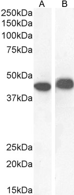
Figure 1. Western blot analysis of AMHR2 using anti-AMHR2 antibody (PB9983). Electrophoresis was performed on a 5-20% SDS-PAGE gel at 70V (Stacking gel) / 90V (Resolving gel) for 2-3 hours. The sample well of each lane was loaded with 30 ug of sample under reducing conditions. Lane 1: rat kidney tissue lysates. Lane 2: rat liver tissue lysates. Lane 3: HEPG2 whole cell lysates. After electrophoresis, proteins were transferred to a nitrocellulose membrane at 150 mA for 50-90 minutes. Blocked the membrane with 5% non-fat milk/TBS for 1.5 hour at RT. The membrane was incubated with rabbit anti-AMHR2 antigen affinity purified polyclonal antibody (Catalog # PB9983) at 0.5 microg/mL overnight at 4°C, then washed with TBS-0.1%Tween 3 times with 5 minutes each and probed with a goat anti-rabbit IgG-HRP secondary antibody at a dilution of 1:5000 for 1.5 hour at RT. The signal is developed using an Enhanced Chemiluminescent detection (ECL) kit (Catalog # EK1002)?with Tanon 5200 system. A specific band was detected for AMHR2 at approximately 42 kDa. The expected band size for AMHR2 is at 42 kDa.
")
")
")
")
Figure 1. Western blot analysis of AMHR2 using anti-AMHR2 antibody (PB9983). Electrophoresis was performed on a 5-20% SDS-PAGE gel at 70V (Stacking gel) / 90V (Resolving gel) for 2-3 hours. The sample well of each lane was loaded with 30 ug of sample under reducing conditions. Lane 1: rat kidney tissue lysates. Lane 2: rat liver tissue lysates. Lane 3: HEPG2 whole cell lysates. After electrophoresis, proteins were transferred to a nitrocellulose membrane at 150 mA for 50-90 minutes. Blocked the membrane with 5% non-fat milk/TBS for 1.5 hour at RT. The membrane was incubated with rabbit anti-AMHR2 antigen affinity purified polyclonal antibody (Catalog # PB9983) at 0.5 microg/mL overnight at 4°C, then washed with TBS-0.1%Tween 3 times with 5 minutes each and probed with a goat anti-rabbit IgG-HRP secondary antibody at a dilution of 1:5000 for 1.5 hour at RT. The signal is developed using an Enhanced Chemiluminescent detection (ECL) kit (Catalog # EK1002)?with Tanon 5200 system. A specific band was detected for AMHR2 at approximately 42 kDa. The expected band size for AMHR2 is at 42 kDa.
Anti-AMACR Antibody Picoband(r)
PB9983-CARRIER-FREE
ApplicationsFlow Cytometry, ImmunoFluorescence, Western Blot, ImmunoCytoChemistry, ImmunoHistoChemistry
Product group Antibodies
ReactivityBovine, Equine, Human, Monkey, Mouse, Rat
TargetAMACR
Overview
- SupplierBoster Bio
- Product NameAnti-AMACR Antibody Picoband(r)
- Delivery Days Customer9
- Application Supplier NoteTested Species: In-house tested species with positive results. Other applications have not been tested. Optimal dilutions should be determined by end users.
- ApplicationsFlow Cytometry, ImmunoFluorescence, Western Blot, ImmunoCytoChemistry, ImmunoHistoChemistry
- CertificationResearch Use Only
- ClonalityPolyclonal
- Concentration500 ug/ml
- Gene ID23600
- Target nameAMACR
- Target descriptionalpha-methylacyl-CoA racemase
- Target synonymsAMACRD, CBAS4, P504S, RACE, RM, alpha-methylacyl-CoA racemase, 2-methylacyl-CoA racemase
- HostRabbit
- IsotypeIgG
- Protein IDQ9UHK6
- Protein NameAlpha-methylacyl-CoA racemase
- Scientific DescriptionBoster Bio Anti-AMACR Antibody Picoband® catalog # PB9983. Tested in Flow Cytometry, IF, IHC, ICC, WB applications. This antibody reacts with Human, Mouse, Rat. The brand Picoband indicates this is a premium antibody that guarantees superior quality, high affinity, and strong signals with minimal background in Western blot applications. Only our best-performing antibodies are designated as Picoband, ensuring unmatched performance.
- ReactivityBovine, Equine, Human, Monkey, Mouse, Rat
- Storage Instruction-20°C,2°C to 8°C
- UNSPSC12352203
Related products
Product group Antibodies
Anti-AMACR AntibodyA82680
ApplicationsWestern Blot, ELISA, ImmunoHistoChemistry
ReactivityHuman
- SizePrice

Product group Antibodies
Anti-AMACR AntibodyAMAB91561
ApplicationsWestern Blot, ImmunoHistoChemistry
ReactivityHuman
TargetAMACR
- SizePrice
Product group Antibodies

AMACR / P504S Antibody (clone 1F1)LS-C764021
ApplicationsImmunoFluorescence, Western Blot, ImmunoHistoChemistry, ImmunoHistoChemistry Paraffin
ReactivityHuman, Mouse, Rat
TargetAMACR
- SizePrice
Product group Antibodies

Goat anti-AMACREB12538
ApplicationsWestern Blot, ELISA
ReactivityHuman
TargetAMACR
- SizePrice
Product group Antibodies
AMACR Monoclonal AntibodyCSB-MA000203
ApplicationsWestern Blot, ELISA, ImmunoHistoChemistry
ReactivityHuman, Mouse, Rat
TargetAMACR
- SizePrice
Product group Antibodies
Amacr Polyclonal AntibodyCAC08104
ApplicationsImmunoFluorescence, Western Blot, ELISA
ReactivityMouse
TargetAMACR
- SizePrice
Product group Antibodies
AMACR antibodyGTX12498
ApplicationsImmunoHistoChemistry, ImmunoHistoChemistry Paraffin
ReactivityHuman
TargetAMACR
- SizePrice