

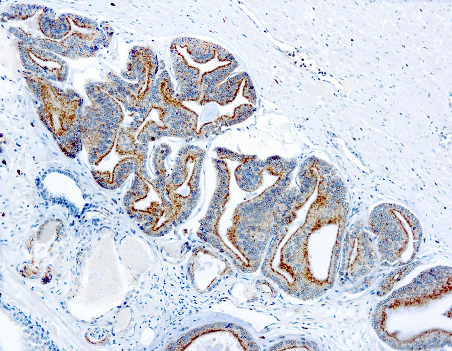
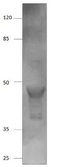
IHC-P analysis of human prostate carcinoma tissue using GTX12498 AMACR antibody.
IHC-P analysis of human prostate carcinoma tissue using GTX12498 AMACR antibody.
AMACR antibody
GTX12498
ApplicationsImmunoHistoChemistry, ImmunoHistoChemistry Paraffin
Product group Antibodies
ReactivityHuman
TargetAMACR
Overview
- SupplierGeneTex
- Product NameAMACR antibody
- Delivery Days Customer9
- Application Supplier NoteIHC-P: 1:25-1:50. *Optimal dilutions/concentrations should be determined by the researcher.Not tested in other applications.
- ApplicationsImmunoHistoChemistry, ImmunoHistoChemistry Paraffin
- CertificationResearch Use Only
- ClonalityPolyclonal
- ConjugateUnconjugated
- Gene ID23600
- Target nameAMACR
- Target descriptionalpha-methylacyl-CoA racemase
- Target synonymsAMACRD, CBAS4, P504S, RACE, RM, alpha-methylacyl-CoA racemase, 2-methylacyl-CoA racemase
- HostRabbit
- IsotypeIgG
- Protein IDQ9UHK6
- Protein NameAlpha-methylacyl-CoA racemase
- Scientific DescriptionThis gene encodes a racemase. The encoded enzyme interconverts pristanoyl-CoA and C27-bile acylCoAs between their (R)- and (S)-stereoisomers. The conversion to the (S)-stereoisomers is necessary for degradation of these substrates by peroxisomal beta-oxidation. Encoded proteins from this locus localize to both mitochondria and peroxisomes. Mutations in this gene may be associated with adult-onset sensorimotor neuropathy, pigmentary retinopathy, and adrenomyeloneuropathy due to defects in bile acid synthesis. Alternatively spliced transcript variants have been described. Read-through transcription also exists between this gene and the upstream neighboring C1QTNF3 (C1q and tumor necrosis factor related protein 3) gene. [provided by RefSeq, Mar 2011]
- ReactivityHuman
- Storage Instruction2°C to 8°C
- UNSPSC41116161
Datasheet
Related products
Product group Antibodies
Anti-AMACR AntibodyA82680
ApplicationsWestern Blot, ELISA, ImmunoHistoChemistry
ReactivityHuman
- SizePrice
Product group Antibodies
Anti-AMACR Antibody144-01130
ApplicationsWestern Blot
ReactivityHuman, Mouse
TargetAMACR
- SizePrice

Product group Antibodies
Anti-AMACR AntibodyAMAB91561
ApplicationsWestern Blot, ImmunoHistoChemistry
ReactivityHuman
TargetAMACR
- SizePrice
Product group Antibodies

AMACR / P504S Antibody (clone 1F1)LS-C764021
ApplicationsImmunoFluorescence, Western Blot, ImmunoHistoChemistry, ImmunoHistoChemistry Paraffin
ReactivityHuman, Mouse, Rat
TargetAMACR
- SizePrice
Product group Antibodies
AMACR Polyclonal AntibodyBS-0840R
ApplicationsWestern Blot, ELISA, ImmunoHistoChemistry, ImmunoHistoChemistry Frozen, ImmunoHistoChemistry Paraffin
ReactivityHuman, Mouse, Rat
TargetAMACR
- SizePrice
Product group Antibodies
AMACR Monoclonal AntibodyCSB-MA000203
ApplicationsWestern Blot, ELISA, ImmunoHistoChemistry
ReactivityHuman, Mouse, Rat
TargetAMACR
- SizePrice
Product group Antibodies

Goat anti-AMACREB12538
ApplicationsWestern Blot, ELISA
ReactivityHuman
TargetAMACR
- SizePrice
Product group Antibodies
Amacr Polyclonal AntibodyCAC08104
ApplicationsImmunoFluorescence, Western Blot, ELISA
ReactivityMouse
TargetAMACR
- SizePrice
Product group Antibodies
AMACR antibodyGTX15893
ApplicationsImmunoFluorescence, ImmunoPrecipitation, Western Blot, ELISA, ImmunoCytoChemistry, ImmunoHistoChemistry, ImmunoHistoChemistry Paraffin
ReactivityHuman, Rat
TargetAMACR
- SizePrice