


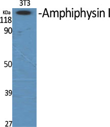
Figure 1. Western blot analysis of Amphiphysin/AMPH using anti-Amphiphysin/AMPH antibody (A02364-1). Electrophoresis was performed on a 5-20% SDS-PAGE gel at 70V (Stacking gel) / 90V (Resolving gel) for 2-3 hours. The sample well of each lane was loaded with 30 ug of sample under reducing conditions. Lane 1: rat brain tissue lysates, Lane 2: mouse brain tissue lysates. After electrophoresis, proteins were transferred to a nitrocellulose membrane at 150 mA for 50-90 minutes. Blocked the membrane with 5% non-fat milk/TBS for 1.5 hour at RT. The membrane was incubated with rabbit anti-Amphiphysin/AMPH antigen affinity purified polyclonal antibody (Catalog # A02364-1) at 0.5 microg/mL overnight at 4°C, then washed with TBS-0.1%Tween 3 times with 5 minutes each and probed with a goat anti-rabbit IgG-HRP secondary antibody at a dilution of 1:5000 for 1.5 hour at RT. The signal is developed using an Enhanced Chemiluminescent detection (ECL) kit (Catalog # EK1002) with Tanon 5200 system. A specific band was detected for Amphiphysin/AMPH at approximately 115-125 kDa. The expected band size for Amphiphysin/AMPH is at 76 kDa.
. Overlay histogram showing U87 cells stained with A02364-1 (Blue line). To facilitate intracellular staining, cells were fixed with 4% paraformaldehyde and permeabilized with permeabilization buffer. The cells were blocked with 10% normal goat serum. And then incubated with rabbit anti-Amphiphysin/AMPH Antibody (A02364-1, 1 microg/1x106 cells) for 30 min at 20°C. DyLight®488 conjugated goat anti-rabbit IgG (BA1127, 5-10 microg/1x106 cells) was used as secondary antibody for 30 minutes at 20°C. Isotype control antibody (Green line) was rabbit IgG (1 microg/1x106) used under the same conditions. Unlabelled sample without incubation with primary antibody and secondary antibody (Red line) was used as a blank control.")
Figure 1. Western blot analysis of Amphiphysin/AMPH using anti-Amphiphysin/AMPH antibody (A02364-1). Electrophoresis was performed on a 5-20% SDS-PAGE gel at 70V (Stacking gel) / 90V (Resolving gel) for 2-3 hours. The sample well of each lane was loaded with 30 ug of sample under reducing conditions. Lane 1: rat brain tissue lysates, Lane 2: mouse brain tissue lysates. After electrophoresis, proteins were transferred to a nitrocellulose membrane at 150 mA for 50-90 minutes. Blocked the membrane with 5% non-fat milk/TBS for 1.5 hour at RT. The membrane was incubated with rabbit anti-Amphiphysin/AMPH antigen affinity purified polyclonal antibody (Catalog # A02364-1) at 0.5 microg/mL overnight at 4°C, then washed with TBS-0.1%Tween 3 times with 5 minutes each and probed with a goat anti-rabbit IgG-HRP secondary antibody at a dilution of 1:5000 for 1.5 hour at RT. The signal is developed using an Enhanced Chemiluminescent detection (ECL) kit (Catalog # EK1002) with Tanon 5200 system. A specific band was detected for Amphiphysin/AMPH at approximately 115-125 kDa. The expected band size for Amphiphysin/AMPH is at 76 kDa.
Anti-Amphiphysin/AMPH Antibody Picoband(r)
A02364-1-BIOTIN
ApplicationsFlow Cytometry, Western Blot, ELISA
Product group Antibodies
ReactivityHuman, Mouse, Rat
TargetAMPH
Overview
- SupplierBoster Bio
- Product NameAnti-Amphiphysin/AMPH Antibody Picoband(r)
- Delivery Days Customer9
- ApplicationsFlow Cytometry, Western Blot, ELISA
- CertificationResearch Use Only
- ClonalityPolyclonal
- Concentration500 ug/ml
- ConjugateBiotin
- Gene ID273
- Target nameAMPH
- Target descriptionamphiphysin
- Target synonymsAMPH1, amphiphysin, amphiphysin (Stiff-Mann syndrome with breast cancer 128kD autoantigen), amphiphysin I
- HostRabbit
- IsotypeIgG
- Protein IDP49418
- Protein NameAmphiphysin
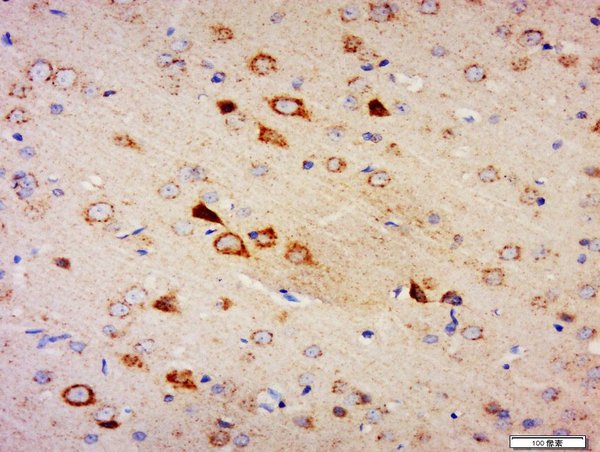
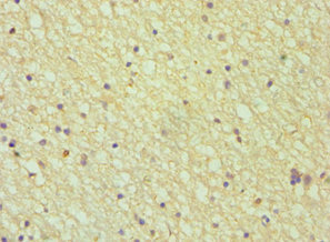
- Scientific DescriptionBoster Bio Anti-Amphiphysin/AMPH Antibody Picoband® catalog # A02364-1. Tested in ELISA, Flow Cytometry, WB applications. This antibody reacts with Human, Mouse, Rat. The brand Picoband indicates this is a premium antibody that guarantees superior quality, high affinity, and strong signals with minimal background in Western blot applications. Only our best-performing antibodies are designated as Picoband, ensuring unmatched performance.
- ReactivityHuman, Mouse, Rat
- Storage Instruction-20°C,2°C to 8°C
- UNSPSC12352203
Related products
Product group Antibodies
Amphiphysin Polyclonal AntibodyBS-2409R
ApplicationsImmunoFluorescence, ELISA, ImmunoCytoChemistry, ImmunoHistoChemistry, ImmunoHistoChemistry Frozen, ImmunoHistoChemistry Paraffin
ReactivityBovine, Canine, Chicken, Equine, Human, Mouse, Rabbit, Rat
TargetAMPH
- SizePrice
Product group Antibodies
ApplicationsImmunoPrecipitation, Western Blot, ImmunoCytoChemistry, ImmunoHistoChemistry
ReactivityMouse, Rat
TargetAMPH
- SizePrice
Product group Antibodies
Anti-AMPH Antibody144-05389
ApplicationsWestern Blot
ReactivityHuman, Mouse, Rat
TargetAMPH
- SizePrice
Product group Antibodies
Anti-AMPH AntibodyA98291
ApplicationsWestern Blot, ELISA
ReactivityHuman, Mouse, Rat
- SizePrice
Product group Antibodies

ApplicationsWestern Blot, ELISA, ImmunoHistoChemistry
ReactivityCanine, Human, Mouse, Rat
TargetAMPH
- SizePrice
Product group Antibodies
Amphiphysin antibody [N1N2], N-termGTX103247
ApplicationsImmunoFluorescence, Western Blot, ImmunoCytoChemistry, ImmunoHistoChemistry, ImmunoHistoChemistry Paraffin
ReactivityHuman, Mouse, Rat
TargetAMPH
- SizePrice
Product group Antibodies

AMPH / Amphiphysin AntibodyLS-C400547
ApplicationsWestern Blot, ELISA, ImmunoHistoChemistry
ReactivityHuman, Mouse
TargetAMPH
- SizePrice

Product group Antibodies
Anti-AMPH AntibodyHPA019828
ApplicationsWestern Blot, ImmunoCytoChemistry, ImmunoHistoChemistry
ReactivityHuman
TargetAMPH
- SizePrice
Product group Antibodies
AMPH AntibodyCSB-PA001683DSR1HU
ApplicationsELISA, ImmunoHistoChemistry
ReactivityHuman
TargetAMPH
- SizePrice