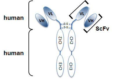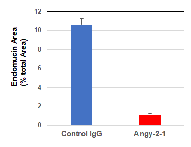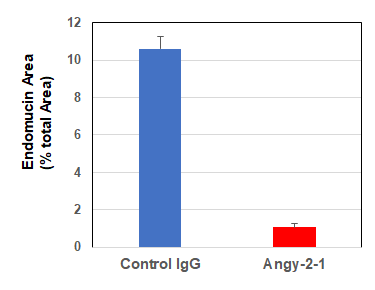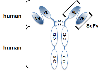
Structure of the recombinant antibody anti-Angiopoietin-2 (human), mAb (rec.) (blocking) (Angy-1-4) (Prod. No. AG-27B-0015). The single chain variable human fragment (ScFv) selected by antibody phage display technology and specific to the antigen of inter
anti-Angiopoietin-2 (human), mAb (rec.) (blocking) (Angy-1-4)
AG-27B-0015
ApplicationsELISA, Neutralisation/Blocking
Product group Antibodies
ReactivityHuman
TargetANGPT2
Overview
- SupplierAdipoGen Life Sciences
- Product Nameanti-Angiopoietin-2 (human), mAb (rec.) (blocking) (Angy-1-4)
- Delivery Days Customer10
- ApplicationsELISA, Neutralisation/Blocking
- CertificationResearch Use Only
- ClonalityMonoclonal
- Clone IDAngy-1-4
- Concentration1 mg/ml
- Estimated Purity>95%
- Gene ID285
- Target nameANGPT2
- Target descriptionangiopoietin 2
- Target synonymsAGPT2, ANG2, LMPHM10, angiopoietin-2, Tie2-ligand, angiopoietin-2B, angiopoietin-2a
- HostHuman
- IsotypeIgG2
- Protein IDO15123
- Protein NameAngiopoietin-2
- Scientific DescriptionAngiopoietin-1 (Ang-1) and Angiopoietin-2 (Ang-2) are closely related secreted ligands which bind with similar affinity to Tie-2. Tie-2 and angiopoietins have been shown to play critical roles in embryogenic angiogenesis and in maintaining the integrity of the adult vasculature. Ang-1 activates Tie-2 signaling on endothelial cells to promote chemotaxis, cell survival, cell sprouting, vessel growth and stabilization. Ang-2 has been identified as a secreted protein ligand of Tie-2 and has alternatively been reported to be an antagonist for Ang-1 induced Tie-2 signaling as well as an agonist for Tie-2 signaling, depending on the cell context. anti-Angiopoietin-2 (human), mAb (rec.) (blocking) (Angy-1-4) is an antibody developed by antibody phage display technology using a human naive antibody gene library. These libraries consist of scFv (single chain fragment variable) composed of VH (variable domain of the human immunoglobulin heavy chain) and VL (variable domain of the human immunoglobulin light chain) connected by a polypeptide linker. The antibody fragments are displayed on the surface of filamentous bacteriophage (M13). This scFv was selected by affinity selection on antigen in a process termed panning. Multiple rounds of panning are performed to enrich for antigen-specific scFv-phage. Monoclonal antibodies are subsequently identified by screening after each round of selection. The selected monoclonal scFv is cloned into an appropriate vector containing a Fc portion of interest and then produced in mammalian cells to generate an IgG like scFv-Fc fusion protein. - Recombinant Antibody. Recognizes human angiopoietin-2. Does not recognize mouse angiopoietin-2 and human angiopoietin-1. Species cross-reactivity: Human. Clone: Angy-1-4. Isotype: Human IgG2lambda. Applications: ELISA, FUNC (Blocking). Host: CHO cells. Liquid. In PBS containing 10% glycerol and 0.02% sodium azide. Angiopoietin-1 (Ang-1) and Angiopoietin-2 (Ang-2) are closely related secreted ligands which bind with similar affinity to Tie-2. Tie-2 and angiopoietins have been shown to play critical roles in embryogenic angiogenesis and in maintaining the integrity of the adult vasculature. Ang-1 activates Tie-2 signaling on endothelial cells to promote chemotaxis, cell survival, cell sprouting, vessel growth and stabilization. Ang-2 has been identified as a secreted protein ligand of Tie-2 and has alternatively been reported to be an antagonist for Ang-1 induced Tie-2 signaling as well as an agonist for Tie-2 signaling, depending on the cell context. anti-Angiopoietin-2 (human), mAb (rec.) (blocking) (Angy-1-4) is an antibody developed by antibody phage display technology using a human naive antibody gene library. These libraries consist of scFv (single chain fragment variable) composed of VH (variable domain of the human immunoglobulin heavy chain) and VL (variable domain of the human immunoglobulin light chain) connected by a polypeptide linker. The antibody fragments are displayed on the surface of filamentous bacteriophage (M13). This scFv was selected by affinity selection on antigen in a process termed panning. Multiple rounds of panning are performed to enrich for antigen-specific scFv-phage. Monoclonal antibodies are subsequently identified by screening after each round of selection. The selected monoclonal scFv is cloned into an appropriate vector containing a Fc portion of interest and then produced in mammalian cells to generate an IgG like scFv-Fc fusion protein.
- ReactivityHuman
- Storage Instruction-20°C,2°C to 8°C
- UNSPSC41116161

