

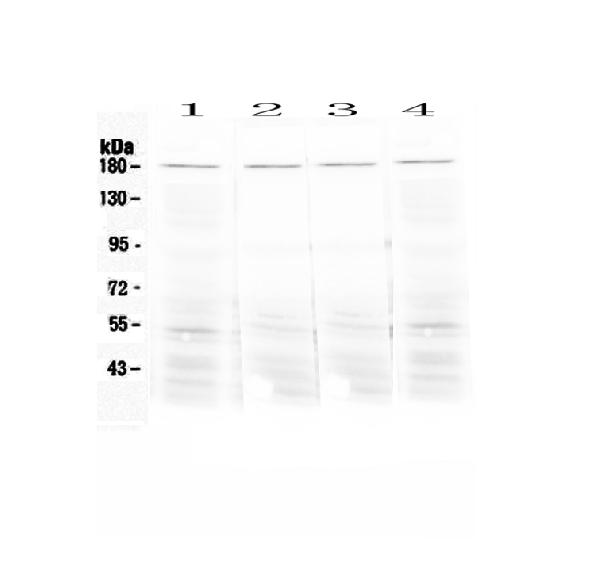
Figure 1. Western blot analysis of Ace using anti-Ace antibody (A00251). Electrophoresis was performed on a 5-20% SDS-PAGE gel at 70V (Stacking gel) / 90V (Resolving gel) for 2-3 hours. The sample well of each lane was loaded with 50ug of sample under reducing conditions. Lane 1: mouse lung tissue lysates, Lane 2: mouse testis tissue lysates, Lane 3: mouse stomach tissue lysates, Lane 4: rat lung tissue lysates. After Electrophoresis, proteins were transferred to a Nitrocellulose membrane at 150mA for 50-90 minutes. Blocked the membrane with 5% Non-fat Milk/ TBS for 1.5 hour at RT. The membrane was incubated with rabbit anti-Ace antigen affinity purified polyclonal antibody (Catalog # A00251) at 0.5 microg/mL overnight at 4°C, then washed with TBS-0.1%Tween 3 times with 5 minutes each and probed with a goat anti-rabbit IgG-HRP secondary antibody at a dilution of 1:10000 for 1.5 hour at RT. The signal is developed using an Enhanced Chemiluminescent detection (ECL) kit (Catalog # EK1002) with Tanon 5200 system. A specific band was detected for Ace at approximately 180KD. The expected band size for Ace is at 150KD.
Figure 1. Western blot analysis of Ace using anti-Ace antibody (A00251). Electrophoresis was performed on a 5-20% SDS-PAGE gel at 70V (Stacking gel) / 90V (Resolving gel) for 2-3 hours. The sample well of each lane was loaded with 50ug of sample under reducing conditions. Lane 1: mouse lung tissue lysates, Lane 2: mouse testis tissue lysates, Lane 3: mouse stomach tissue lysates, Lane 4: rat lung tissue lysates. After Electrophoresis, proteins were transferred to a Nitrocellulose membrane at 150mA for 50-90 minutes. Blocked the membrane with 5% Non-fat Milk/ TBS for 1.5 hour at RT. The membrane was incubated with rabbit anti-Ace antigen affinity purified polyclonal antibody (Catalog # A00251) at 0.5 microg/mL overnight at 4°C, then washed with TBS-0.1%Tween 3 times with 5 minutes each and probed with a goat anti-rabbit IgG-HRP secondary antibody at a dilution of 1:10000 for 1.5 hour at RT. The signal is developed using an Enhanced Chemiluminescent detection (ECL) kit (Catalog # EK1002) with Tanon 5200 system. A specific band was detected for Ace at approximately 180KD. The expected band size for Ace is at 150KD.
Anti-Angiotensin Converting Enzyme 1/Ace Antibody Picoband(r)
A00251-DYLIGHT594
ApplicationsFlow Cytometry, Western Blot, ImmunoHistoChemistry
Product group Antibodies
ReactivityMouse, Rat
TargetAce
Overview
- SupplierBoster Bio
- Product NameAnti-Angiotensin Converting Enzyme 1/Ace Antibody Picoband(r)
- Delivery Days Customer9
- ApplicationsFlow Cytometry, Western Blot, ImmunoHistoChemistry
- CertificationResearch Use Only
- ClonalityPolyclonal
- Concentration500 ug/ml
- ConjugateOther Conjugate
- Gene ID11421
- Target nameAce
- Target descriptionangiotensin I converting enzyme
- Target synonymsCD143, angiotensin-converting enzyme, angiotensin I converting enzyme (peptidyl-dipeptidase A) 1, dipeptidyl carboxypeptidase I, dipeptidyl peptidase, kininase II
- HostRabbit
- IsotypeIgG
- Protein IDP09470
- Protein NameAngiotensin-converting enzyme
- Scientific DescriptionBoster Bio Anti-Angiotensin Converting Enzyme 1/Ace Antibody Picoband® catalog # A00251. Tested in Flow Cytometry, IHC, WB applications. This antibody reacts with Mouse, Rat. The brand Picoband indicates this is a premium antibody that guarantees superior quality, high affinity, and strong signals with minimal background in Western blot applications. Only our best-performing antibodies are designated as Picoband, ensuring unmatched performance.
- ReactivityMouse, Rat
- Storage Instruction-20°C,2°C to 8°C
- UNSPSC12352203
Related products
Product group Antibodies
ApplicationsImmunoPrecipitation, Western Blot, ImmunoCytoChemistry, ImmunoHistoChemistry
ReactivityMouse
TargetAce
- SizePrice
Product group Antibodies
ACE antibody [9B21]GTX53158
ApplicationsWestern Blot
ReactivityMouse
TargetAce
- SizePrice
Product group Antibodies
Anti-Angiotensin Converting Enzyme 1/Ace Antibody Picoband(r)A00251-CARRIER-FREE
ApplicationsFlow Cytometry, Western Blot, ImmunoHistoChemistry
ReactivityMouse, Rat
TargetAce
- SizePrice