


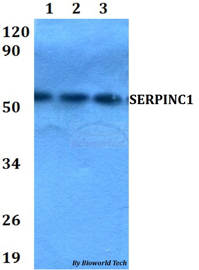
Figure 1. Western blot analysis of Antithrombin III/SERPINC1 using anti-Antithrombin III/SERPINC1 antibody (A00469-1). Electrophoresis was performed on a 5-20% SDS-PAGE gel at 70V (Stacking gel) / 90V (Resolving gel) for 2-3 hours. The sample well of each lane was loaded with 30 ug of sample under reducing conditions. Lane 1: rat liver tissue lysates, Lane 2: mouse testis tissue lysates, Lane 3: mouse ovary tissue lysates, Lane 4: mouse liver tissue lysates. After electrophoresis, proteins were transferred to a nitrocellulose membrane at 150 mA for 50-90 minutes. Blocked the membrane with 5% non-fat milk/TBS for 1.5 hour at RT. The membrane was incubated with rabbit anti-Antithrombin III/SERPINC1 antigen affinity purified polyclonal antibody (Catalog # A00469-1) at 0.5 microg/mL overnight at 4°C, then washed with TBS-0.1%Tween 3 times with 5 minutes each and probed with a goat anti-rabbit IgG-HRP secondary antibody at a dilution of 1:5000 for 1.5 hour at RT. The signal is developed using an Enhanced Chemiluminescent detection (ECL) kit (Catalog # EK1002) with Tanon 5200 system. A specific band was detected for Antithrombin III/SERPINC1 at approximately 53 kDa. The expected band size for Antithrombin III/SERPINC1 is at 52 kDa.
. Overlay histogram showing HepG2 cells stained with A00469-1 (Blue line). The cells were fixed with 4% paraformaldehyde and blocked with 10% normal goat serum. And then incubated with rabbit anti-Antithrombin III/SERPINC1 Antibody (A00469-1, 1 microg/1x106 cells) for 30 min at 20°C. DyLight®488 conjugated goat anti-rabbit IgG (BA1127, 5-10 microg/1x106 cells) was used as secondary antibody for 30 minutes at 20°C. Isotype control antibody (Green line) was rabbit IgG (1 microg/1x106) used under the same conditions. Unlabelled sample without incubation with primary antibody and secondary antibody (Red line) was used as a blank control.")
Figure 1. Western blot analysis of Antithrombin III/SERPINC1 using anti-Antithrombin III/SERPINC1 antibody (A00469-1). Electrophoresis was performed on a 5-20% SDS-PAGE gel at 70V (Stacking gel) / 90V (Resolving gel) for 2-3 hours. The sample well of each lane was loaded with 30 ug of sample under reducing conditions. Lane 1: rat liver tissue lysates, Lane 2: mouse testis tissue lysates, Lane 3: mouse ovary tissue lysates, Lane 4: mouse liver tissue lysates. After electrophoresis, proteins were transferred to a nitrocellulose membrane at 150 mA for 50-90 minutes. Blocked the membrane with 5% non-fat milk/TBS for 1.5 hour at RT. The membrane was incubated with rabbit anti-Antithrombin III/SERPINC1 antigen affinity purified polyclonal antibody (Catalog # A00469-1) at 0.5 microg/mL overnight at 4°C, then washed with TBS-0.1%Tween 3 times with 5 minutes each and probed with a goat anti-rabbit IgG-HRP secondary antibody at a dilution of 1:5000 for 1.5 hour at RT. The signal is developed using an Enhanced Chemiluminescent detection (ECL) kit (Catalog # EK1002) with Tanon 5200 system. A specific band was detected for Antithrombin III/SERPINC1 at approximately 53 kDa. The expected band size for Antithrombin III/SERPINC1 is at 52 kDa.
Anti-Antithrombin III/SERPINC1 Antibody Picoband(r)
A00469-1-CARRIER-FREE
ApplicationsFlow Cytometry, Western Blot, ELISA
Product group Antibodies
ReactivityHuman, Mouse, Rat
TargetSERPINC1
Overview
- SupplierBoster Bio
- Product NameAnti-Antithrombin III/SERPINC1 Antibody Picoband(r)
- Delivery Days Customer9
- ApplicationsFlow Cytometry, Western Blot, ELISA
- CertificationResearch Use Only
- ClonalityPolyclonal
- Concentration500 ug/ml
- Gene ID462
- Target nameSERPINC1
- Target descriptionserpin family C member 1
- Target synonymsAT3, AT3D, ATIII, ATIII-R2, ATIII-T1, ATIII-T2, THPH7, antithrombin-III, serine (or cysteine) proteinase inhibitor, clade C (antithrombin), member 1, serpin peptidase inhibitor clade C member 1, serpin peptidase inhibitor, clade C (antithrombin), member 1
- HostRabbit
- IsotypeIgG
- Protein IDP01008
- Protein NameAntithrombin-III
- Scientific DescriptionBoster Bio Anti-Antithrombin III/SERPINC1 Antibody Picoband® catalog # A00469-1. Tested in ELISA, Flow Cytometry, WB applications. This antibody reacts with Human, Mouse, Rat. The brand Picoband indicates this is a premium antibody that guarantees superior quality, high affinity, and strong signals with minimal background in Western blot applications. Only our best-performing antibodies are designated as Picoband, ensuring unmatched performance.
- ReactivityHuman, Mouse, Rat
- Storage Instruction-20°C,2°C to 8°C
- UNSPSC12352203
Related products
Product group Antibodies
Anti-SERPINC1 AntibodyA28836
ApplicationsWestern Blot
ReactivityHuman, Mouse, Rat
- SizePrice
Product group Antibodies
Anti-SERPINC1 Antibody144-01574
ApplicationsImmunoFluorescence, Western Blot, ImmunoHistoChemistry
ReactivityHuman, Mouse
TargetSERPINC1
- SizePrice
Product group Antibodies
SERPINC1 Recombinant Antibody, Biotin ConjugatedBSM-61553R-BIOTIN
ApplicationsWestern Blot
ReactivityHuman
TargetSERPINC1
- SizePrice
Product group Antibodies
SERPINC1 Polyclonal AntibodyCAC14879
ApplicationsWestern Blot, ELISA, ImmunoHistoChemistry
ReactivityRat
TargetSERPINC1
- SizePrice
Product group Antibodies
SERPINC1 AntibodyCSB-PA021079LA01HU
ApplicationsWestern Blot, ELISA, ImmunoHistoChemistry
ReactivityHuman, Rat
TargetSERPINC1
- SizePrice

Product group Antibodies

Antithrombin-III AntibodyLS-C331551
ApplicationsImmunoFluorescence, Western Blot, ImmunoHistoChemistry
ReactivityHuman, Mouse
TargetSERPINC1
- SizePrice

Product group Antibodies
Anti-SERPINC1 AntibodyHPA001816
ApplicationsImmunoHistoChemistry
ReactivityHuman
TargetSERPINC1
- SizePrice
Product group Antibodies
Antithrombin III antibodyGTX113132
ApplicationsImmunoFluorescence, Western Blot, ImmunoCytoChemistry, ImmunoHistoChemistry, ImmunoHistoChemistry Paraffin
ReactivityHuman
TargetSERPINC1
- SizePrice