


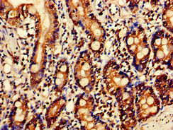
Immunohistochemical staining of human bone marrow shows strong cytoplasmic positivity in bone marrow poietic cells.
Immunohistochemical staining of human bone marrow shows strong cytoplasmic positivity in bone marrow poietic cells.
Anti-SERPINC1 Antibody
HPA001816
ApplicationsImmunoHistoChemistry
Product group Antibodies
ReactivityHuman
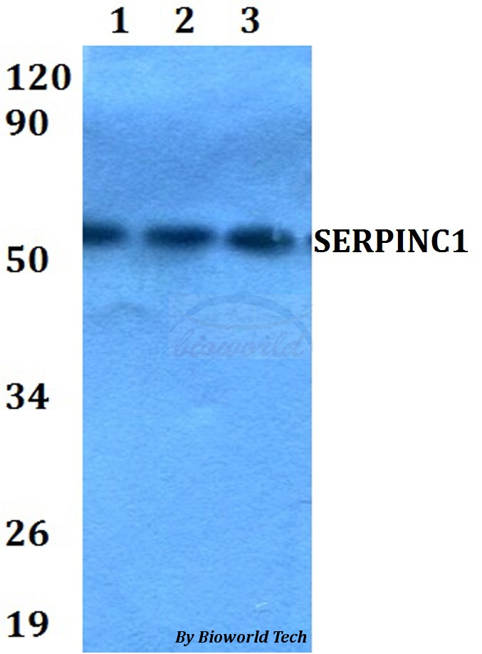
TargetSERPINC1
Overview
- SupplierAtlas Antibodies
- Product NameAnti-SERPINC1 Antibody
- Delivery Days Customer4
- ApplicationsImmunoHistoChemistry
- CertificationResearch Use Only
- ClonalityPolyclonal
- ConjugateUnconjugated
- Gene ID462
- Target nameSERPINC1
- Target descriptionserpin family C member 1
- Target synonymsAT3, AT3D, ATIII, ATIII-R2, ATIII-T1, ATIII-T2, THPH7, antithrombin-III, serine (or cysteine) proteinase inhibitor, clade C (antithrombin), member 1, serpin peptidase inhibitor clade C member 1, serpin peptidase inhibitor, clade C (antithrombin), member 1
- HostRabbit
- IsotypeIgG
- Protein IDP01008
- Protein NameAntithrombin-III
- Scientific DescriptionRecombinant Protein Epitope Signature Tag (PrEST) antigen sequence
- ReactivityHuman
- Storage Instruction-20°C,2°C to 8°C
- UNSPSC41116161
Datasheet
MSDS
Related products
Product group Antibodies
Anti-SERPINC1 AntibodyA28836
ApplicationsWestern Blot
ReactivityHuman, Mouse, Rat
- SizePrice
Product group Antibodies
Anti-SERPINC1 Antibody144-01574
ApplicationsImmunoFluorescence, Western Blot, ImmunoHistoChemistry
ReactivityHuman, Mouse
TargetSERPINC1
- SizePrice

Product group Antibodies
Anti-Antithrombin III/SERPINC1 Antibody Picoband(r)A00469-1-CARRIER-FREE
ApplicationsFlow Cytometry, Western Blot, ELISA
ReactivityHuman, Mouse, Rat
TargetSERPINC1
- SizePrice
Product group Antibodies
SERPINC1 Recombinant Antibody, Biotin ConjugatedBSM-61553R-BIOTIN
ApplicationsWestern Blot
ReactivityHuman
TargetSERPINC1
- SizePrice
Product group Antibodies
SERPINC1 Polyclonal AntibodyCAC14879
ApplicationsWestern Blot, ELISA, ImmunoHistoChemistry
ReactivityRat
TargetSERPINC1
- SizePrice
Product group Antibodies
SERPINC1 AntibodyCSB-PA021079LA01HU
ApplicationsWestern Blot, ELISA, ImmunoHistoChemistry
ReactivityHuman, Rat
TargetSERPINC1
- SizePrice
